
Before you read the answer you will probably want to review the original post of the mystery quiz from last week.
The Answer:
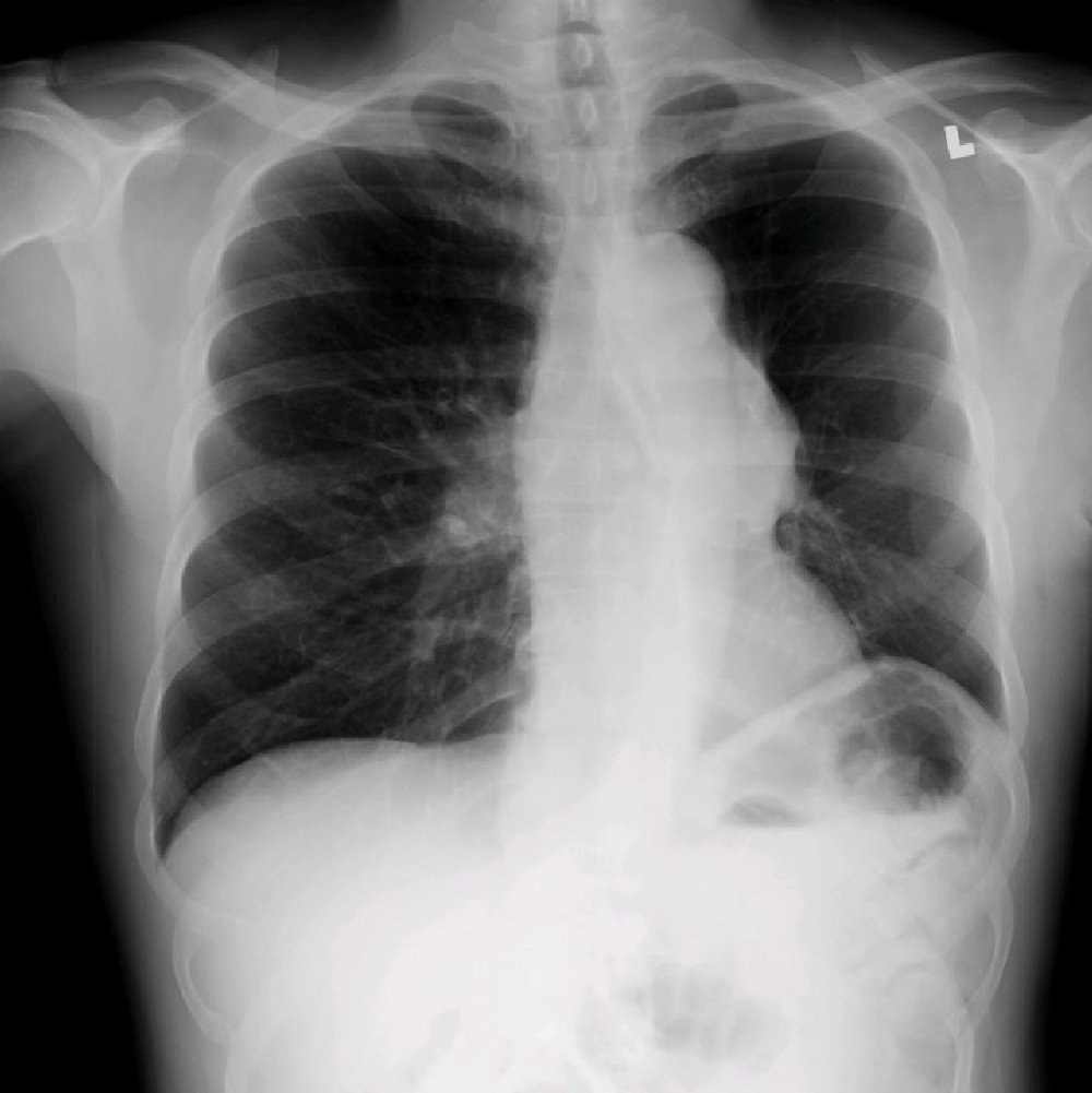
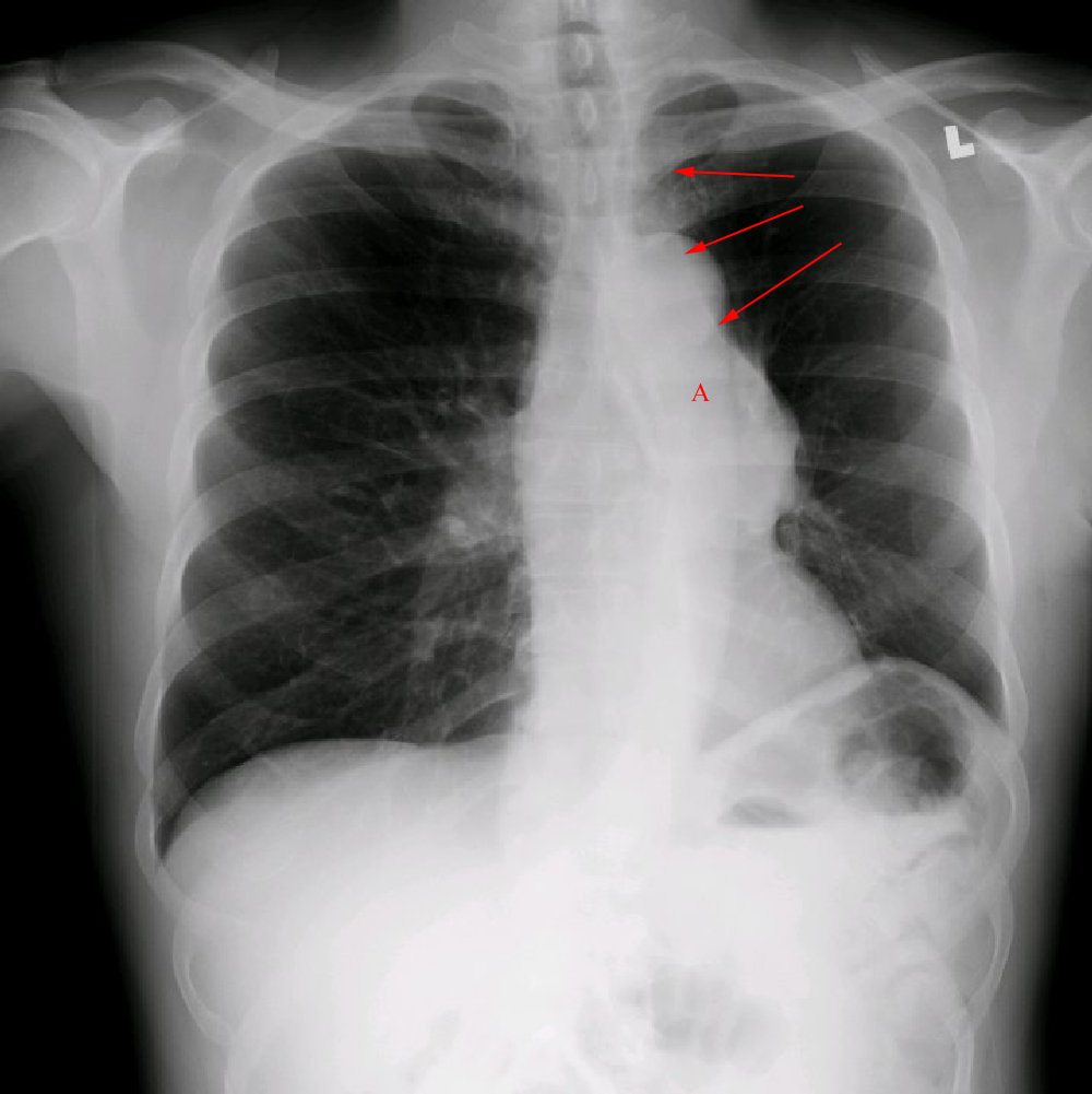
The chest film shows a probable mass in the area of the left hilum and associated complete collapse of the left upper lobe. The key findings are loss of volume of the left hemithorax indicated by elevation of the left hemidiaphragm and shift of the mediastinum to the left side. Additionally, the arrows indicate the major fissure, ordinarily not visible, but now bordering the left upper lobe because it has shifted upward. The increased density seen at the left hilum (A)suggests the presence of a mass. Click on the thumbnails below.
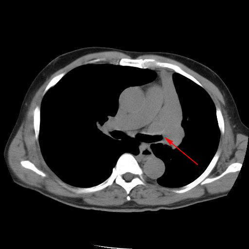
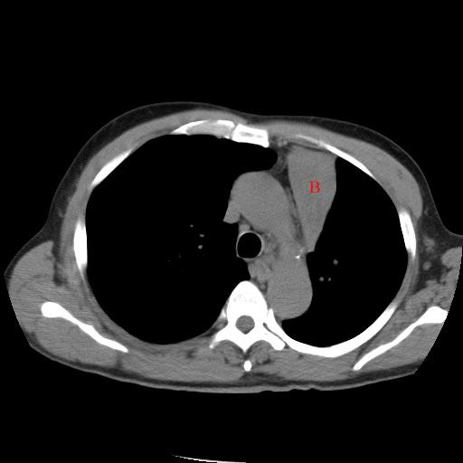
All of the above findings are more easily seen on the two CT images below. Note the cutoff of the left upper lobe bronchus by an intraluminal mass (arrow), and the airlessness of the collapsed left upper lobe (B).
This patient subsequently developed hoarseness related to involvement of the left recurrent laryngeal nerve which was invaded by tumor thus indicating non-resectability of this lesion. The biopsy of the endobronchial lesion at the left upper lobe revealed non-small cell carcinoma.
Although a peanut or foreign body was not responsible for these findings, several respondents correctly identified the volume loss and implied collapse of the left upper lobe.
2 comments on “Mystery Quiz #1-The Answer…”
Forever humbled.
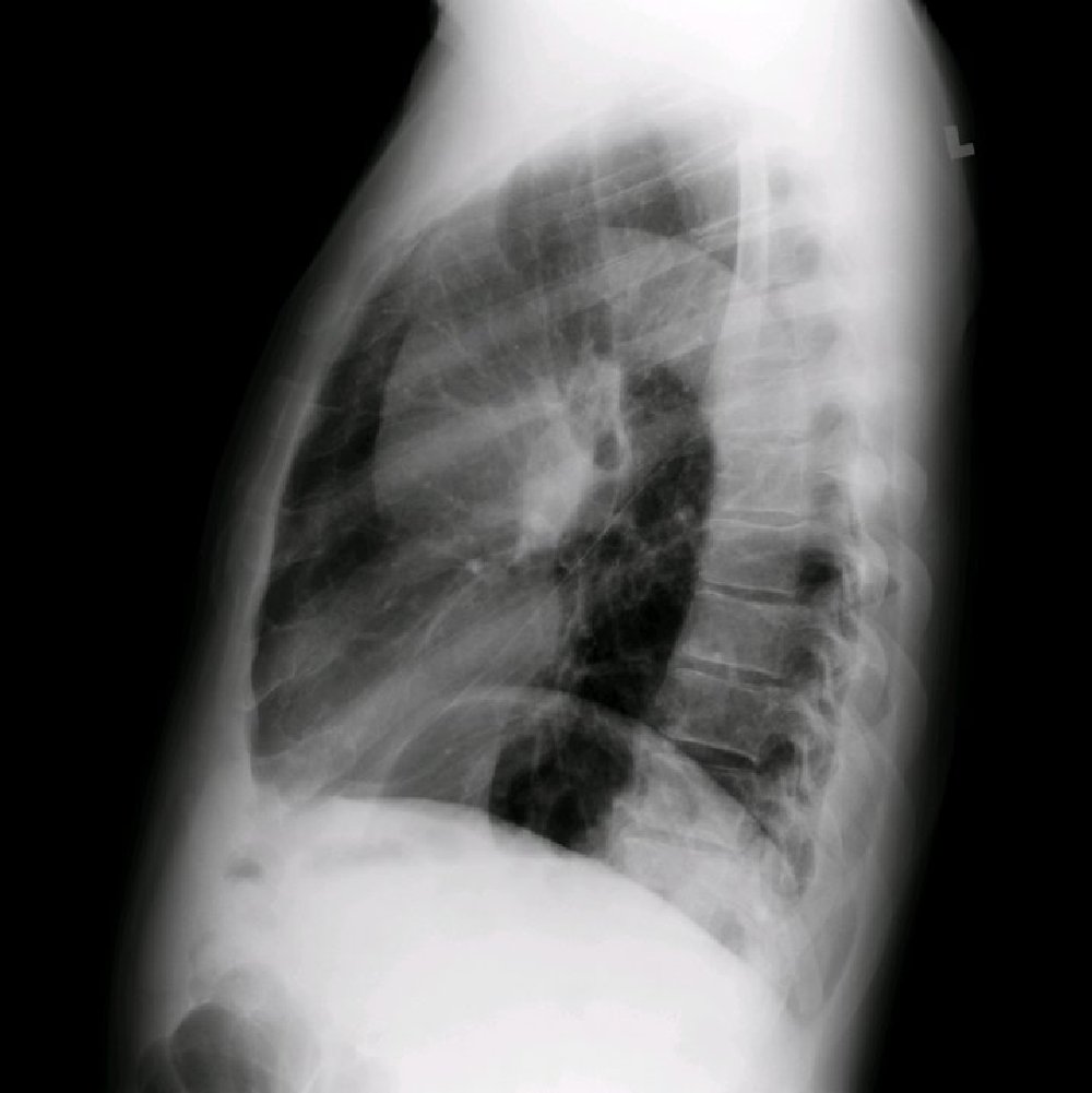
what’s the wedge-shaped density seen on the lateral XR over part of the heart?
I guess hyporaemia in the LUL was imaginary
The wedge-shaped density overlying the heart is a small anterior hernia (Morgagni)…
Comments are closed.