Faculty Peer Reviewed
Mr. R is a 46-year-old man with a past medical history of polycythemia vera on hydroxyurea and chronic hepatitis B and C who presented with acutely worsening left upper-quadrant abdominal pain. This occurred in the context of 3 months of worsening abdominal pain and 1.5 years of increasing abdominal distension. His physical exam was remarkable for massive splenomegaly (18cm span) and a non-palpable liver.
Laboratory findings are significant for microcytic anemia with an elevated RDW, thrombocytopenia, elevated PT, PTT, and INR, elevated D-dimer, normal AFP, and normal liver enzymes. CT of the abdomen and pelvis, MRV, and MRI suggested chronic portal vein thrombosis with cavernous transformation, splenic vein thrombosis, massive splenomegaly, esophageal varices, and liver cirrhosis. No focal liver or splenic lesions were noted. Polycythemia vera is a clonal disorder of multipotent hematopoietic progenitor cells, and is characterized by the accumulation of phenotypically normal red blood cells, granulocytes, and platelets in the absence of a recognizable physiologic stimulus. As the most common chronic myeloproliferative disorder, polycythemia vera presents in patients as an elevated hemoglobin or hematocrit, and can be further defined with laboratory evaluation as an increase in red cell mass. The definition of polycythemia  vera as a disease of panmyelosis encompasses both its primary features and complications, including splenomegaly, pruritis, erythromyalgia, bleeding, and hypercoagulability with arterial and venous thrombosis.
In 2005 a chromosomal clue to the molecular basis of polycythemia vera was elucidated. Certain patients with myeloproliferative disorders, including polycythemia vera, essential thrombocythemia, and myelofibrosis, can manifest a loss of heterozygosity on the short arm of chromosome 9. Subsequent microsatellite mapping of a cohort of these patients identified a somatic, dominant, gain-of-function mutation in a region that included the pseudokinase domain of the JAK2 gene. The JAK2 V617F mutation was then described as a base pair transversion that results in a single amino acid substitution of phenylalanine for valine on the short arm of chromosome 9.1
Not all patients with polycythemia vera have a loss of heterozygosity at the 9p locus; rather, this 9p locus merely identifies the location of the JAK2 allele, and patients with polycythemia may be heterozygous or homozygous for the JAK2 V617F mutation. It is the JAK2 V617F mutation, however, regardless of its homozygous or heterozygous presentation on a chromosomal level, that is seen in almost all polycythemia vera patients. Over 95% of patients with this myeloproliferative disorder have this mutation in one of these two forms; the other 5% have other JAK2 mutations with functional consequences similar to those noted for JAK2V617F.2 The JAK2 mutation, therefore, is virtually pathogneumonic for polycythemia vera.
JAK2 functions as a cytoplasm tyrosine kinase that transduces signals from hematopoietic growth factors in both normal and euplastic multipotent hematopoietic progenitor cells. Because the characteristic JAK2 mutation occurs in hematopoietic progenitor cells, the circulating erythrocytes, granulocytes, and platelets that derive from these euplastic precursors also harbor the mutation. This results in a panmyelosis, with marked qualitative and quantitative dysfunction that affects all three lineages.
Polycythemia vera, however, is classically characterized by neoplastic erythroid cells that proliferate in the absence of erythropoietin because of constitutive signaling by the mutated JAK2 protein. While this results in the increased red blood cell number and mass that are part of the diagnostic criteria for polycythemia vera, this hyperviscosity is not the sole mechanism of the hypercoagulability that also seen in the disease. While high whole blood viscosity due to high hematocrit certainly contributes to the hypercoagulable state of polycythemia vera, phlebotomy reduces but does not eliminate the increased risk of thrombotic events. Hence, hypercoagulability in polycythemia vera is a multifactorial process.
Hypercoagulable states include both primary inherited and secondary acquired disorders associated with increased risks of thromboembolism. While the mechanism of hypercoagulability in these inherited states, such as factor V Leiden, antithrombin III mutation, and protein C or S deficiency, can be summarized as abnormal regulation of the coagulation cascade, the pathophysiology of increased thrombotic events in the acquired disorders is more variable. Several heterogeneous disease states, including cancer, myeloproliferative disorders, antiphospholipid antibody syndrome, and heparin-induced thrombocytopathy may predispose to thrombosis; the mechanisms of hypercoagulability in these diseases, however, differ vastly.
What is especially intriguing about the myeloproliferative disorders, and specifically polycythemia pera, is that the same mutation that defines the neoplastic, clonal proliferation of tri-lineage myeloid precursors in the bone marrow and their descendants in the blood is also responsible for the hypercoagulable state of the disease.  This JAK2 mutation of polycythemia vera not only defines the molecular basis for the disease, but is also responsible for its sequelae, including increased red blood cell number and mass, hypercoagulability, and pruritis, as well as its response to hydroxyurea, the current mainstay of therapy.
Although the myeloproliferative disorders have been linked to splanchnic venous thrombosis, such as Budd-Chiari syndrome and portal vein thrombosis for many years, the mechanism of hypercoagulability has only recently been elucidated.
Early research, later proven to be incorrect, proposed that splanchnic veno-occlusive disease was caused by direct damage to sinusoidal endothelial cells in the portal circulation, resulting in local embolism by sinusoidal lining cells and subsequent circulatory blockage.3 Subsequent studies hypothesized roles for decreased natural anticoagulants, increased plasma homocysteine, anticardiolipin antibodies, or inherited thrombophilic genotypes (such as factor V Leiden, prothrombin mutation, and protein C deficiency) in promoting arterial disease and splanchnic venous thrombosis in patients with polycythemia vera.
Although portal and hepatic venous thrombosis are most often the result of more than one thrombotic risk factor and are not due to single, isolated events,4 thrombophilic genotypes or deficient functional natural anticoagulants are equally prevalent among polycythemia vera patients with arterial disease, splanchnic venous thrombosis, or no thrombotic events at all.5 These observations suggested that an additional process – one that was independent of the classically inherited mechanisms that increase the risk of thromboembolism -was responsible for the hypercoagulable state of polycythemia vera.
The hypercoagulable state of polycythemia vera is the direct result of the JAK2 mutation. As a consequence of mutated JAK2 function, there is a generalized hypersensitivity to cytokines, with over-expression of procoagulant factors and adhesion molecules at the vascular wall. Specifically, there is activation of hemostasis, with increased expression of platelet-associated tissue factor microparticles and resultant increased formation of platelet-neutrophil aggregates.6 The JAK2 mutation also results in platelet hypersensitivity to these prothrombotic signals, as they undergo spontaneous activation, product secretion (i.e., thromboxane A2), and aggregation mediated by von Willebrand factor. Platelet plugs then transiently occlude the microvascular circulation, deaggregate, and recirculate in a defective form.7 The quantitative and qualitative dysfunctions of platelets in polycythemia vera, as well as the hypersensitivity to cytokines at the level of the endothelial wall, explain the hypercoagulable state of the disease.
The defective circulating platelets also, however, explain the bleeding diathesis that is another, seemingly paradoxical, characteristic of the disease. Leukocytosis, due to hyperproliferation from JAK2 kinase activity, also contributes to increased thrombotic risk and cardiovascular endpoints. White blood cell counts over 15 x 109 are positively correlated with increased risk for cardiovascular death, stroke, transient ischemic attack, myocardial infarction, peripheral arterial thrombosis, deep venous thrombosis of the leg or abdominal veins, and pulmonary embolism.8 While the molecular mechanism for this increased thrombotic risk due to leukocytosis has yet to be definitively elucidated, one may hypothesize that increased neutrophil numbers promote enhanced interactions with transient platelet plugs and a prothrombotic endothelial wall, thereby increasing the probability of significant vascular occlusive events.
The JAK2 mutation is also likely responsible for the intense pruritis that did not affect Mr. R, but that affects up to 85% of polycythemia vera patients. Granulocytosis, a component of the JAK2 mutation-associated panmyelosis, results in increased circulating numbers of both basophils and mast cells. Although no formal mechanism has been described for basophil hyperactivation and subsequent degranulation, mast cells of polycythemia patients are not only more numerous, but also functionally different from those of normal individuals. Mast cells from polycythemia vera patients are more resistant to apoptosis and release quantitatively more puritogenic cytokines than do normal mast cells.9 The sequelae of polycythemia vera, therefore, including hypercoagulability and pruritis, are direct results of the JAK2 mutation; it induces both panmyelosis and hypersensitivity of increased circulating numbers of erythrocytes, platelets, and granulocytes to cytokines.
It is perhaps only fitting, therefore, that the JAK2 mutation also predicts the response of patients with polycythemia vera to chemotherapy. The presence of the JAK2 mutation and, specifically, its allele burden, is inversely correlated with the daily dose of hydroxyurea needed for adequate bone marrow suppression. Hydroxyurea inactivates ribonucleotide reductase, thereby inhibiting DNA synthesis and DNA repair, and inducing cell death in S phase. Higher allele burdens of the JAK2 mutation required smaller daily doses of hydroxyurea, suggesting that the antiproliferative effect of hydroxyurea is most potent in patients with higher levels of JAK2-associated stem cell myeloproliferation.10 The presence of this JAK2 mutation in multipotent hematopoietic stem cells and their tri-lineage descendants dictates the molecular mechanism, hyperviscosity, hypercoagulability, pruritis, and current treatment approach to polycythemia vera.
It is tempting to speculate, however, about how the therapeutic approach to patients like Mr. R. will evolve in the future. With this focused, well-defined target providing a source for most, if not all, manifestations of polycythemia vera, perhaps specific targeting of the pathophysiologic effects of the V617F chromosomal mutation and its resultant gain-of-function JAK2 cytoplasmic tyrosine kinase may alleviate the manifestations of this myeloproliferative disease. Small molecule inhibitors of JAK2 are currently under development and in early phase clinical trials.11 While the results of these studies may be neither timely nor conclusive enough to alter the therapy or the course of Mr. R.’s disease, they will take the first step toward investigating the clinical efficacy of JAK2 inhibitors, both specifically in patients with polycythemia vera and more generally in those with other myeloproliferative disorders.
Commentary By Bruce Raphael, MD, Clinical Professor of Medicine, Division of Hematology NYU Langone Medical Center:
This a very good review of the complications of polycythemia vera and the proposed molecular basis of the disease with its consequent changes in numbers and function of all the hematopoietic cells derived from a stem cell that has a JAK2 mutation. This mutation, unlike many hematologic malignancies with complex cytogenetic and molecular changes, would appear to be solely responsible for the disease state. That said, the same mutation can manifest as essential thrombocythemia while other patients in the heterozygote or homozygous state have similar presentation as p. vera despite the different allelic load. Hence, there is probably a host cellular response to the mutation that affects the phenotypic presentation. The hope that inhibition of the JAK2 derived tyrosine kinase will cure this disease as in BCR/abl positive CML has not been evident in the early studies further complicating the hypothesis of JAK2 driven hematopoiesis.
Emily Slater is a 4th year medical student at NYU School of Medicine
Peer reviewed by Bruce Raphael, MD, Clinical Professor, Department of Medicine Division of Hematology, NYU Langone Medical Center
References:
1.  Kralovics R. et al. “A gain-of-function mutation of JAK2 in myeloproliferative disorders.â€Â NEJM 352(2005): 1779-1790.
2.  Tefferi, A. “Polycythemia Vera – molecular mechanisms and clinical applications.â€Â NEJM 356(2007): 444-445.
3.  Poreddy V. and DeLeve, L.D. “Hepatic circulatory diseases associated with chronic myeloid disorders.â€Â Clinics in Liver Disease 6(2002): 909-931.
4.  Denninger M.H., Chait Y., Casadevall N., et al. “Cause of portal or hepatic venous thrombosis in adults: the role of multiple concurrent factors.â€Â Hepatology 31(2000): 153-159.
5.  Amitrano, L. et al. “Thrombophilic genotypes, natural anticoagulants, and plasma homocysteine in myeloproliferative disorders: relationship with splanchnic venous thrombosis and arterial disease.â€Â American Journal of Hematology 72(2003): 75-81.
6.  Falanga A., Barbui T., and Rickles, F.R. “Hypercoagulability and tissue factor upregulation in hematologic malignancies.â€Â Seminars in Thrombosis and Hemostasis: Tissue Factor and Cancer 34(2008): 204-210.
7.  Michiels J.J. et al. “The paradox of platelet activation and impaired function: platelet-von Willebrand factor interactions, and the etiology of thrombotic and hemorrhagic manifestations in Essential Thrombocythemia and Polycythemia Vera.â€Â Semin Thromb Hemost 32(2006): 589-604.
8 . Landolfi R. et al. “Leukocytosis as a major thrombotic risk factor in patients with Polycythemia Vera.â€Â Blood 109(2007): 2446-2452.
9.  Mesa, R.A. “Itchy mast cells in myeloproliferative neoplasms.â€Â Inside Blood 113(2009): 5697-5698.
10.  Sirhan S. et al. “The presence of JAK2V617F in primary myelofibrosis or its allele burden in Polycythemia Vera predicts chemosensitivity to Hydroxyurea.â€Â Hematology 83(2008): 363-365.
11.  Pardanani A. “JAK2 inhibitor therapy in myeloproliferative disorders: rationale, preclinical studies and ongoing clinical trials.â€Â Leukemia 22(2008): 23-30.
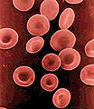
Image courtesy of Wikimedia Commons.


One comment on “Polycythemia Vera Presenting as a Hypercoagulable State: What is the Pathophysiologic Role of JAK2 in the Mechanism, Manifestations, and Treatment of the Disease?”
Bruce,
Great review. Kudos also to yourmedical student.
Doing better,
Tom
Comments are closed.