 Commentary by Judith Brenner MD, Associate Program Director, NYU Internal Medicine Residency Program
A 66 year old woman with a history of dyslipidemia and remote tobacco use presents with a sudden onset of pain located in her posterior left thigh radiating down her left leg below the knee. The pain began during the course of an upper respiratory illness with a cough. The pain is burning in quality and is bothersome day and night. NSAIDs have been taken and relieve the pain temporarily.
What is Lasegue’s sign and when would you find it useful?
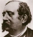
“Lasegue’s sign” is another name for the straight leg maneuver used in the diagnosis of lumbosacral radiculopathy. It is named after the French clinician, Charles Lasegue, whose student described the maneuver and named it after his mentor. To perform the straight leg test, the clinician lifts the extended leg of a patient in a supine position. A positive response occurs when the pain pattern of the lumbar radiculopathy is reproduced. The test should be stopped when the pain is reproduced or maximum flexion is achieved. It is believed that the pain is reproduced because of stretching of the sciatic nerve and its roots when the leg is flexed. The sensitivity and specificity of the maneuver is reported in McGee’s Evidence Based Physical Diagnosis as 73-98% and 11-61%, respectively. More useful than the straight leg maneuver is the crossed straight leg test. This test has a lower sensitivity but a specificity that approaches 90%. The crossed straight leg maneuver is performed by raising the unaffected leg in a similar manner to the straight leg test. The examiner looks for the reproduction of radicular pain with elevation of the opposite leg. This test is reported by McGee to have a higher specificity and, thus, a higher likelihood ratio (LR=4.3).
The differential diagnosis of a positive straight leg test includes: disc protrusion with impingement of nerve roots below L4; meningismus; any intraspinal lesion such as tumor below L4; malignant disease or osteomyelitis of the ilium or upper femur; ankylosing spondylitis; fractured sacrum and more.
Other signs to help in the diagnosis of a lumbar radiculopathy are:
• Weak ankle dorsiflexion: LR of 4.9
• Ipsilateral calf wasting: LR 5.2
• Abnormal ankle jerk: LR 4.3
• Crossed straight leg maneuver: LR 4.3
In the case presented, the patient reports the sudden onset of radicular pain in a setting where acute disc herniation is likely. On exam, findings of any of the above mentioned signs would make the diagnosis more likely. In order to localize further, one would perform a sensory exam or a more comprehensive motor exam.
In this case, the patient did indeed have a disc herniation at L5/S1, verified on MRI. She was treated with analgesics, including Tylenol #3 for 3 weeks, and began physical therapy as her pain began to remit. Her symptoms fully resolved within 8 weeks.
Excellent references:
Deyo RA. Low Back Pain: Review Article. NEJM 2001;344:363
Deyo RA. What can the History and Physical Exam Tell us about LBP. JAMA 1992;268:760.

2 comments on “Bedside Rounds: What is Lasegue’s Sign?”
Excellent idea to have such a forum.
If the Lasegue Test is negative, but the patient has still experienced the classic symptons of Sciatica, ( a source of pain which seems to emanate from a point some inches to the left of the lower spine, radiating into the buttock, down the back of the thigh etc) is this a sign of a torn muscle and if so, what then is the best treatment ?
Comments are closed.