
 Commentary by Bani Chander MD, PGY-3. and Ben Bergman MD, PGY-3
Please also see last week’s Class Act Post on the pathogenesis of Rheumatic Heart DiseaseÂ
A 34-year-old Hispanic male presents with the chief complaint of chest pain. The patient had been well until 10 days prior to admission, when he developed a severe sore throat accompanied by fever, rigors, and diffuse myalgias. There was no associated cough. Three days later, he visited his primary care physician at an outside facility and was prescribed penicillin 500 mg twice daily for presumed streptococcal pharyngitis. Five days later, he presented to the emergency room of an outside hospital and stopped his antibiotics after a throat culture there was negative for beta-hemolytic strep. The following morning, the patient awoke in the middle of the night with severe left-sided chest pain and presented again to his local emergency room. The patient described a sensation of left-sided chest pressure which became worse when lying down and somewhat improved upon sitting up.
An electrocardiogram was done and revealed ~2mm ST segment elevations in II, III, aVF, and V3-V6; the initial troponin was 14.7. A diagnosis of an ST-elevation myocardial infarction was made and alteplase was administered for thrombolysis. He was also given intravenous unfractionated heparin, aspirin, clopidogrel, metopolol, simvastatin, and sublingual nitroglycerin. The patient had transient relief of his chest pain after these medications but the pain recurred a few hours later. Given his persistent chest pain and ST segment elevations despite thrombolytics, he was transferred to Bellevue for cardiac catheterization.
Upon admission to the CCU, the patient was febrile to 100.8â—¦ and was still having persistent chest pain but had no associated dyspnea, palpitations, nausea, vomiting, or diaphoresis. An electrocardiogram showed persistent 2 mm ST elevations in leads II, aVF, and V3-V6 with diffuse PR segment depression and a normal PR interval. On physical examination, he was a well-appearing male in no distress. He had enlarged tonsils but no exudates; a single 0.5 cm tender left anterior cervical lymph node was palpable. There was no jugulovenous distension. The cardiac exam did not reveal any murmurs, rubs, or gallops, and the PMI was non-displaced. His pulmonary, musculoskeletal, dermatologic, and neurologic exams were all unremarkable.
The patient underwent cardiac catheterization on the first day of admission. The coronary arteries were angiographically normal. The anteroapical, apical, and inferoapical walls were hypokinetic, with an estimated ejection fraction of 45 %. The patient was continued on aspirin, clopidogrel, and metoprolol and started on lisinopril and simvastatin. A cardiac MRI was performed, revealing inflammation of the myopericardium, decreased left ventricular systolic function, and no significant valvular regurgitation or lesions. Transthoracic echocardiography showed a normal ejection fraction with apical and inferior wall hypokinesis, with mild mitral and tricuspid insufficiency.
Laboratory testing was remarkable for a leukocyte count of 27.6 and a troponin of 12.7. A serum anti-streptolysin-O antibody was sent and returned as 1541 IU (normal < 200). Tests for echovirus, parvovirus, and coxsackie virus were all negative. Hepatitis serologies and HIV testing were also negative. His TSH was normal at 1.9. Epstein-Barr virus IgM was negative, while IgG was positive. The erythrocyte sedimentation rate was 48 and the C-reactive protein was 83. Over the hospital course, the ASO titer increased to 2982 IU. The patient was started on penicillin G and high-dose aspirin for presumed acute rheumatic fever. After two days, the patient developed mild epigastric pain despite the concurrent use of a proton-pump inhibitor, and high-dose aspirin was discontinued. A repeat transthoracic echocardiogram 5 days after the initial TTE showed improved left ventricular wall motion, and the patient was discharged home with instructions to receive monthly intramuscular injections of penicillin G benzathine 1.2 million units for five to ten years. Prior to his discharge, contact with his outside primary care physician confirmed that his initial culture prior to starting antibiotics was positive for group A beta-hemolytic streptococcus.
Discussion:
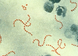
Acute rheumatic fever (ARF) is a delayed, non-suppurative sequela of infection with group A beta-hemolytic streptococci or Streptococcus pyogenes. The acute phase of ARF is caused by a proliferative inflammatory reaction that involves the connective and/or collagen tissues and commonly affects the heart, joints, brain and subcutaneous tissues, although any organ may be involved.1
The concept of “rheumatogenicity†came from the observation that only certain strains of Group A streptococcus were associated with acute rheumatic fever. The observed strains include the M serotypes 1, 3, 5, 6, 14, 18, 24, 27, and 29; however, given the genetic diversity among strains, any group A streptococcus may acquire the potential to cause acute rheumatic fever.2 An estimated 60% of cases of acute rheumatic fever lead to rheumatic heart disease, which carries an annual mortality of 1.5% without medical treatment.3 Worldwide it is estimated that the prevalence of RHD is at least 15-20 million with 233,000 deaths per year.
In 1944, Duckett Jones published his famous “Jones Criteria,†which in its updated form is the basis of the current approach to acute rheumatic fever. The diagnosis is made with evidence of a preceding group A streptococcal (GAS) infection (either positive culture or rapid streptococcal antigen test or elevated or rising streptococcal antibody titer) with the presence of two major or one major and two minor criteria (see table 1). The measurement of antistreptolysin-O antibody has become the definitive biochemical test to document the presence of antecedent GAS infection. In patients with acute infection due to GAS, the ASO antibody titer can be elevated after one week, reaching maximal levels in 3 to 5 weeks, and in the absence of re-infection or treatment failure typically falls to pre-infection levels in 6 to 12 months.5,6
Acute rheumatic fever is generally considered to be a disease of the young, although varying age ranges have been published, with case reports of the initial syndrome in adults as old as 38 years. The introduction of antibiotics has lead to a rapid decline in the incidence of ARF and in the United States the incidence following a streptococcal pharyngitis infection has decreased from 100 per 100,000 people at the beginning of the 20th century, to less than 2 per 100,000 at the present time.7 Worldwide the incidence is estimated at 0.23-1.88 patients per 100,000, and in children of Polynesian ancestry in Hawaiian and Maori populations, the incidence is still up to 13.4 patients per 100,000 hospitalized children per year.8
Table 1: The Revised Jones Criteria9
Major criteria
Carditis
Polyarthritis
Chorea
Erythema marginatum
Subcutaneous nodules
Minor criteria
Clinical
Arthralgia
Fever
Laboratory
Elevated erythrocyte sedimentation rate, C reactive protein
EKG
First degree atrioventricular block
Treatment of GAS infection has resulted in a dramatic decrease in the incidence of rheumatic heart disease in the United States. The disease persists with significantly higher incidence and morbidity in many countries with less access to diagnostic testing and antibiotics. While the management of rheumatic heart disease and the prevention of rheumatic fever are key medical successes of the twentieth century, active rheumatic carditis remains poorly understood and relatively untreatable.
There are few proven therapies for acute rheumatic fever and the treatment of rheumatic carditis is even more limited. Carditis occurs in ~50% of patients with ARF and may involve the pericardium, myocardium, or the valve cusps. It can cause permanent damage to the heart valves and may result in death even years after the initial presentation of carditis. This particular patient was treated in the hospital with penicillin G in order to treat the underlying streptococcal infection; however, given that rheumatic carditis is presumed to be an inflammatory disease, we conducted several investigations looking into therapies for treatment including steroids, immunoglobulin, and salicylates.
A recent Cochrane review of treatment with steroids or immunoglobulin for patients with rheumatic carditis failed to show a reduction in cardiac complications at one year.10 Treatment with salicylates and steroids in acute rheumatic fever still remains relatively controversial. Use of high dose salicylates has been studied and there is some evidence that they may reduce symptoms acutely; however, there is no improvement in long-term clinical or cardiac outcomes.11,12 Some physicians recommend treatment with aspirin at a dose of 75 mg/kg/day; however, this dose has only been studied in children and there are no available data with adults. In the US-UK trial of 49 patients aged 16 or less with acute rheumatic fever, treatment with a 12-week course of salicylates versus bed rest showed no apparent difference between them either during their hospital stay or at five- year follow-up. Not only was treatment with salicylates ineffective in controlling disease activity, but it may have been potentially harmful.11 The suggested daily aspirin dose of 75 mg/kg resulted in gastritis in our patient. Data are limited regarding the use of salicylates other than aspirin.
Ultimately, although the prevalence of rheumatic heart disease is significantly lower in recent years, the implications of treatment for active rheumatic carditis on the development of valvulitis and subsequent valvular sequelae still remain poorly understood and relatively undefined.