Vivian Hayashi MD and Robert Smith MD, Mystery Quiz Section Editors
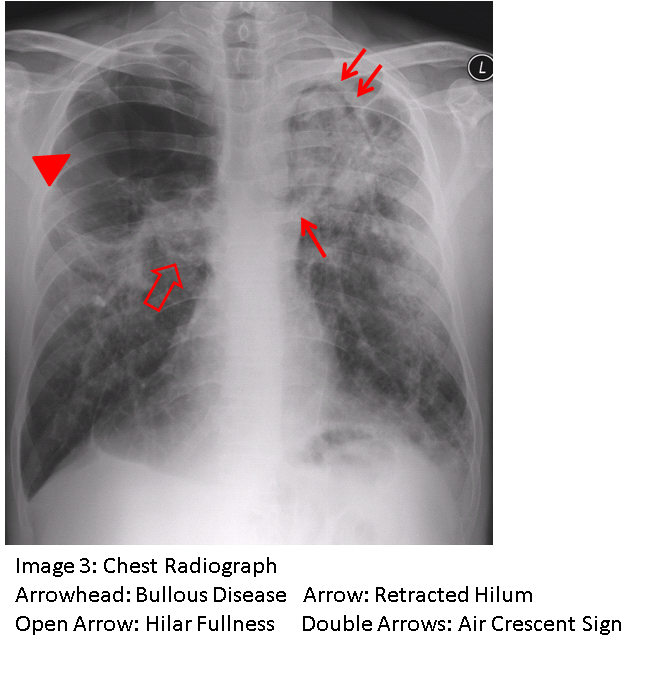
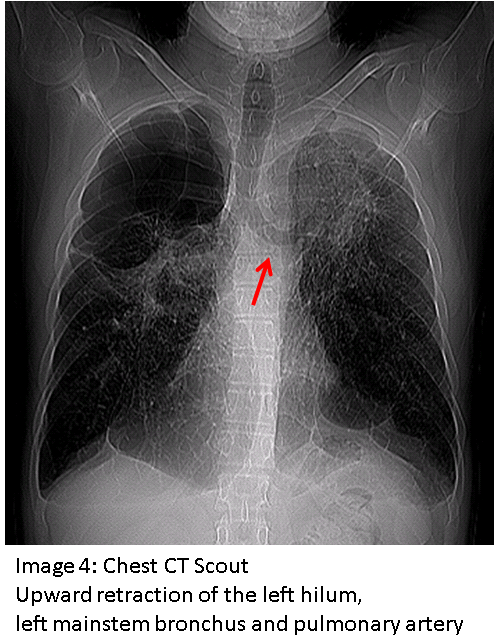
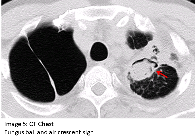
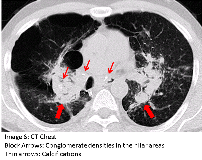
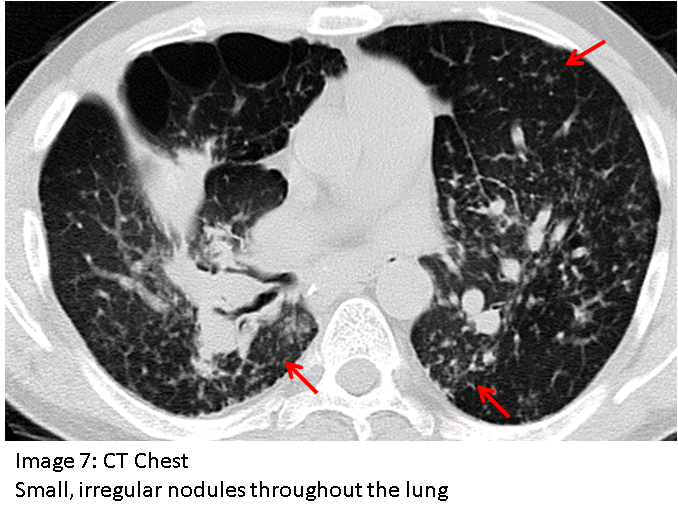
The answer to the mystery quiz is progressive massive fibrosis. This condition is a severe form of silicosis. The chest radiograph (Image 3) shows bullous disease of the right upper lobe, increased density of the right hilum, increased density and upward retraction of the left hilum (also seen in Image 4), increased density in peripheral areas of both lungs, and an air crescent sign in the left upper lobe (also seen in Image 5). The air crescent sign indicates the presence of a fungus ball (aspergilloma ) contained within a cavity. Image 6 shows conglomerate densities of both hila (block arrows) with calcification (thin arrows). Image 7 shows profuse small, irregular nodules throughout the lung.
Silicosis results from the inhalation of crystalline silica which is silicon dioxide (SiO2). This compound is abundantly present in rocks, such as sandstone, granite and slate. Occupations which involve blasting, mining, or tunneling in rock may expose workers to inhalation of silica. Workers in ceramics and glass manufacturing can also be exposed to inhalable silica. Silicosis is categorized as simple silicosis when pulmonary nodules are less than 10mm in size. This condition usually affects the upper lung zones and often manifests as innumerable nodules. Patients may be asymptomatic at this stage. Usually, the disease appears 10 to 30 years after exposure. When present before this time, as in our case, the disease is called accelerated silicosis as opposed to chronic silicosis. When the nodules of simple silicosis coalesce to form large conglomerate densities on imaging (Image 6), the disease is classified as progressive massive fibrosis. This condition, which is more frequently associated with accelerated silicosis, distorts the lung and results in areas of emphysema which are adjacent to areas of dense conglomerate fibrosis. The conglomerate densities may cavitate, sometimes spontaneously but often due to mycobacterial infection. Our patient was infected with Mycobacterium avium complex. Silicosis is believed to arise from injury due to oxygen free radicals, and ongoing toxicity may persist even after an individual is no longer exposed to silica.
Our case illustrates the multiple complications of progressive massive fibrosis, including emphysema and pulmonary cavitation associated with aspergilloma and atypical mycobacterial infection. Although these complications were listed as individual entries in the differential diagnosis of the mystery quiz, progressive massive fibrosis best explains all of the findings in the case. The treatment of our patient included frequent courses of antibiotics, anti-mycobacterial medications, supplemental oxygen, and bronchodilator therapy with occasional courses of prednisone. Other than lung transplantation, treatment options are limited.

