
 By Christopher Sonne, MD
Peer reviewed
Learning Objectives:
1. Understand the pathophysiology of peritonitis secondary to bowel perforation.
2. Understand how classic findings of peritonitis can be absent in some cases of bowel perforation.
3. Recognize bowel perforation as a complication requiring emergent surgical intervention.
Case:
An 89 year-old woman with a history of chronic constipation was admitted for several weeks of severe constipation and abdominal bloating. On initial exam, she exhibited abdominal distention but had minimal abdominal tenderness. Initial CT imaging demonstrated a significant stool burden with prominent colonic distension extending from the cecum to the transverse colon. She had no flatus and required nasogastric tube decompression, with several days of laxatives, scheduled enemas, and attempts at manual disimpaction. Conservative management was unsuccessful at relieving her constipation. Frequent enemas and an aggressive bowel regimen continued.
On hospital day 6, the patient complained of a dull left groin pain. Serial abdominal exams continued to be unremarkable. However, after several hours she became increasingly lethargic and hypotensive. At this time, physical examination revealed crepitus extending from the left groin to below the left knee despite an abdominal exam which continued to lack rigidity, tenderness, or guarding. Repeat CT imaging revealed subcutaneous and deep thigh soft tissue emphysema extending from the retroperitoneum likely originating from a colonic perforation. An emergent surgery consultation was made; however, the patient and family elected not to pursue surgical intervention, opting instead for comfort measures. The patient expired early the next morning.
Discussion:
In the practice of clinical inpatient medicine, there can be an over-reliance on waiting for the development of a “surgical abdomen†before seeking an urgent surgical evaluation. This case demonstrates one example of a medicine patient with an uncommon presentation of a common surgical emergency — a colonic perforation presenting without classic abdominal peritoneal signs. Colonic perforation can occur in various conditions treated on the medicine wards including but not limited to diverticulosis, colonic neoplasms, inflammatory bowel disease, stercoral perforations in elderly patients, and direct instrumentation of the colonic mucosa in procedures such as colonoscopy. Some medications such as NSAIDS and steroids have also been associated with an increased incidence of diverticular perforations [1].
When the colon perforates, it spills colonic contents into surrounding sterile anatomical compartments. In contrast to spontaneous bacterial peritonitis which is commonly monomicrobial, secondary peritonitis is predictably polymicrobial. A small perforation releases more than 400 unique bacterial species into the surrounding space [2]. Very quickly, this number is reduced to several dominant species. Escherichia coli and Bacteroides fragilis are most common and have been shown to grow synergistically in their new extra-colonic environment [3]. The bacteria and their accompanying endotoxins promote the influx of complement and both innate and adaptive immune cells, with the subsequent production of pro-inflammatory cytokines TNF-α, IL-1, and IL-6 [4].
If the colon perforates into the intraperitoneal space, rising concentrations of inflammatory cytokines in the intraperitoneal cavity activate mesothelial and mast cells on the visceral peritoneal surface, increasing capillary permeability and resulting in peritoneal edema [4]. Visceral peritoneal nociceptors then signal the afferent visceral nerves, arising from the T8-T10 spinal roots, which produce colicky, poorly-differentiated abdominal pain [5]. As the underlying visceral peritoneum becomes further inflamed, nociceptors on the overlying parietal peritoneum signal well-localizing somatic nerves creating the sharp pain recognizable as the classic peritoneal signs: tenderness on palpation and movement, guarding, and rebound tenderness. If the infection is not controlled, cellular production of nitric oxide, the “terminal mediator” of the inflammatory response begins to block cellular respiration leading to cell death, microvascular injury, acidosis and eventual organ failure [4].
This progression of symptoms related to intraperitoneal inflammation, for example, is evident in a typical presentation of appendicitis. In early stages of appendicitis, the visceral innervation of the appendix causes the patient to experience poorly-localized abdominal pain that generally refers to the peri-umbilical region. As the appendix becomes further inflamed and edematous, the poorly localized pain worsens as the appendix expands into contact with the visceral peritoneum, also innervated by visceral nerves. However, the patient’s pain eventually evolves into severe, localized, right lower quadrant pain as the inflammatory process spreads outward to the overlying somatically innervated parietal peritoneum. Patients with this characteristic evolution of symptoms are often spared further diagnostic workup in favor of immediate surgical intervention before the appendix perforates.
However, there are exceptions to this typical progression of symptoms after an intestinal perforation, as seen in the case above. An understanding of colonic anatomy is key to understanding alternate presentations of a colonic perforation.
The colon is both an intra- and extra-peritoneal organ, depending on its various segments. The transverse colon and sigmoid colon are intraperitoneal with the visceral peritoneum completely covering all external surfaces of the bowel. The ascending colon on the right and the descending colon on the left have their posterior and lateral walls embedded in the retroperitoneum, and their anterior and medial walls covered by visceral peritoneum [6]. As the sigmoid colon continues from its intraperitoneal location towards the rectum, there exists a peritoneal reflection external to the middle third of the rectum. This peritoneal reflection ends the intraperitoneal space, and the remaining rectum exists in the extraperitoneal pelvis. The peritoneal reflection correlates roughly with the anatomic level of the middle rectal valve, or valve of Houston [7].
When it comes to colonic perforations, the transition zones between intraperitoneal and extraperitoneal segments are the most vulnerable to perforation by instrumentation. The transitions between ileum and cecum, and between the distal descending and sigmoid colons, are the most common sites of iatrogenic perforations during colonoscopy [8]. Though rare, rectal perforations distal to the middle valve of Houston leak intrarectal contents and air into the extraperitoneal pelvis. Air travels down pressure gradients along fascial planes, potentially causing pneumoscrotum and subcutaneous emphysema of the lower extremities. In the case above, it is believed that repeated insertions of a plastic enema nozzle may have led to the rectal perforation that resulted in the patient’s deep tissue thigh emphysema.
If a perforation occurs in a retroperitoneal colonic segment, retroperitoneal air and contaminants can travel widely via communicating fascial planes [9,10,11]. Caudally, a plane along the transversalis fascia provides a communication between the retroperitoneum and the extraperitoneal pelvis which again can cause pneumoscrotum and subcutaneous emphysema [12]. Cranially, retroperitoneal air can also travel via endothoracic-endoabdominal fascial planes which communicate with the mediastinum causing pneumomediastinum and subcutaneous crepitus in the chest and neck [9,10].
In the presence of suspected bowel perforation, either with intraperitoneal or extraperitoneal findings, CT imaging of the abdomen with water soluble contrast is most often the diagnostic test of choice if the patient is stable enough to undergo the study. CT imaging of areas with crepitus, if present, will also help to establish the presence of subcutaneous air [9].
Bowel perforation is typically a surgical emergency; resection of the perforated bowel is needed to gain source control of evolving sepsis. Broad-spectrum antibiotics should be started to cover common intracolonic organisms. Markers of systemic inflammation such as tachycardia, hypotension, fevers, elevated lactate, and leukocytosis, along with exam findings of perforation may lend credence to taking a patient to the operating room without further workup.
Conclusion:
Colonic perforation is a potential complication of numerous conditions treated on the medical floor, and requires the emergent assessment for possible surgical intervention. For patients who are known to be at risk for a perforation, there may be an over-reliance on focusing solely on the development of a “surgical abdomen†as a sign of perforation. The case above illustrates extra-peritoneal findings of a colonic perforation in a patient with a benign abdominal exam.
Dr. Christopher Sonne, internal medicine resident at NYU Langone Health
Peer reviewed by Andrew Dikman, MD, Gastroenterology, NYU Langone Health
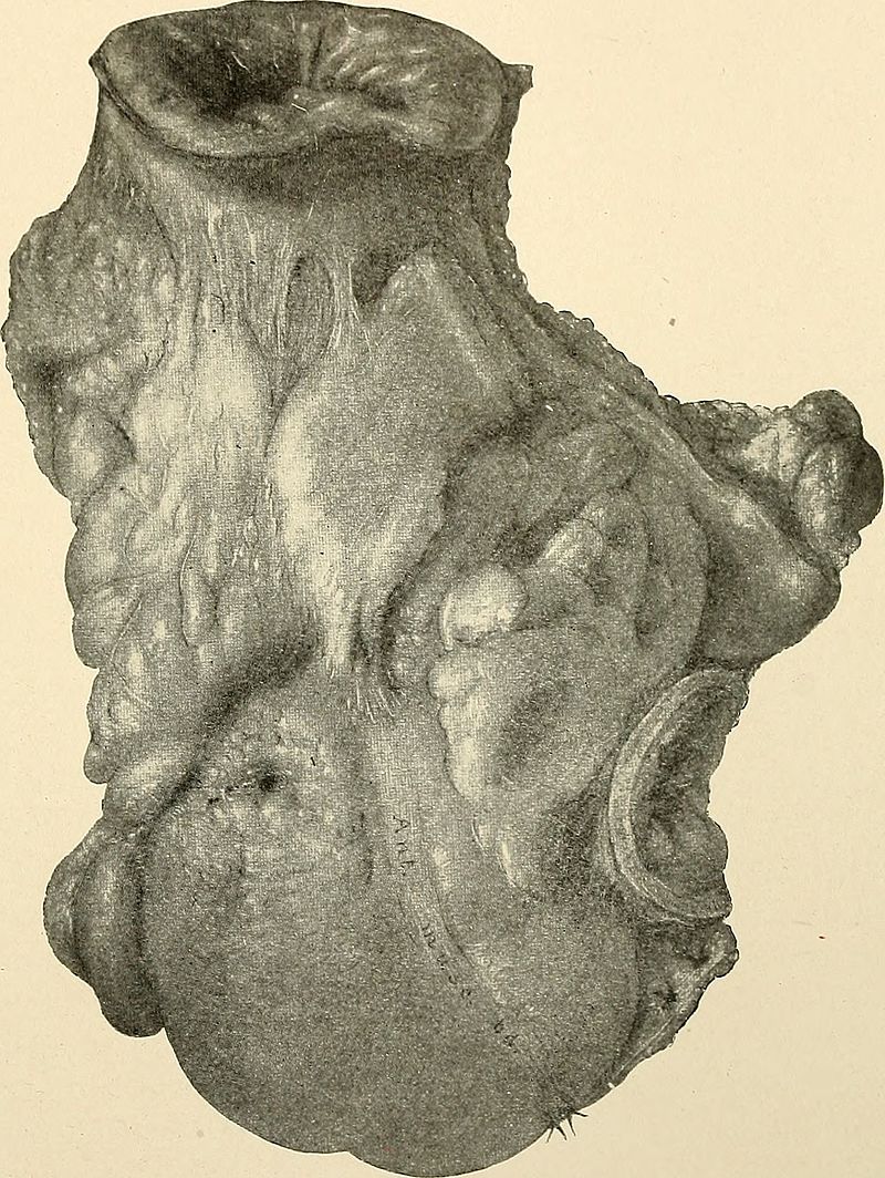
Image courtesy of Wikimedia Commons
References
1. Morris, C. R., et al. “Antiâ€inflammatory drugs, analgesics and the risk of perforated colonic diverticular disease.” British journal of surgery 90.10 (2003): 1267-1272.  https://www.ncbi.nlm.nih.gov/pubmed/14515298
2. Wittmann, Dietmar H., Moshe Schein, and Robert E. Condon. “Management of secondary peritonitis.” Annals of surgery 224.1 (1996): 10. https://www.researchgate.net/profile/Dietmar_Wittmann/publication/14522371_327_Management_of_Secondary_Peritonitis/links/570577fc08aef745f7176cb5/327-Management-of-Secondary-Peritonitis.pdf?origin=publication_list
3. Bartlett, John G., et al. “A review: lessons from an animal model of intra-abdominal sepsis.” Archives of Surgery 113.7 (1978): 853-857.
4. Shein, Moshe, et al. “Hypothesis: compartmentalization of cytokines in intraabdominal infection.” Surgery 119.6 (1996): 694-700. https://www.ncbi.nlm.nih.gov/pubmed/8650611
5. Jänig, Wilfrid. “Neurobiology of visceral afferent neurons: neuroanatomy, functions, organ regulations and sensations.” Biological psychology 42.1 (1996): 29-51.
6. Cappell MS. Large Bowel Disorders. In: McKean SC, Ross JJ, Dressler DD, Scheurer DB. eds. Principles and Practice of Hospital Medicine, 2e New York, NY: McGraw-Hill; . http://accessmedicine.mhmedical.com.ezproxy.med.nyu.edu/content.aspx?bookid=1872§ionid=146982683. Accessed September 16, 2017.
7. Netter, Frank Henry, and Sharon Colacino. Atlas of human anatomy. Plate 371. Ciba-Geigy Corporation, 1989.
8. Putcha, Rajesh V., and J. Steven Burdick. “Management of iatrogenic perforation.” Gastroenterology Clinics of North America 32.4 (2003): 1289-1309. https://europepmc.org/abstract/med/14696308
9. Lee, Mathew John, and Tara M. Connelly. “Head and neck subcutaneous emphysema, a rare complication of iatrogenic perforation during colonoscopy: management review of reported cases from 2000-2016.” Expert Review of Gastroenterology & Hepatology 11.9 (2017): 849-856.
10. Yang, Jun, et al. “Pneumothorax, pneumomediastinum, pneumoperitoneum and extensive subcutaneous emphysema resulting from endoscopic mucosal resection secondary to colonoscopy: A case report.” Oncology letters 11.4 (2016): 2763-2767.
11. Denadai, Rafael, et al. “Rectal perforation after colonoscopic polypectomy presented as subcutaneous emphysema, pneumomediastinum and pneumoretroperitoneum successfully treated conservatively in an elderly adult.” Journal of the American Geriatrics Society 61.8 (2013): 1433-1435.
12. Vilaça, Ana Frias, Alcinda M. Reis, and Isabel M. Vidal. “The anatomical compartments and their connections as demonstrated by ectopic air.” Insights into imaging 4.6 (2013): 759-772.  https://www.ncbi.nlm.nih.gov/pmc/articles/PMC3846937/