
Before you read the answer you should read the orginal post form last week
The Final Poll Results (26 votes): metastatic disease (26%) , mycobacterial disease (22%) ,fungal disease (22%), bronchiolitis obliterans with organizing pneumonia (boop) (13%), septic emboli (9%) ,vasculitis, e.g. wegener’s (4%), thromboembolic disease (4%), sarcoid (0%)
The patient had granulomatous inflammation on pathology with acid-fast organisms seen. The culture grew mycobacterium avium (MAC). After treatment with azithromycin, ethambutol and rifabutin for eighteen months, the follow-up imaging showed significant clearing of the infiltrates.
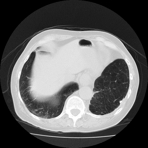
This case highlights one of the imaging patterns that are associated with MAC infection, that is, mostly nodular appearing infiltrates without a predilection for the upper lobes, as seen with reactivation MTB. What was unusual in this case was the rapidity of the disease progression. In other regards, the presentation was typical: mild symptoms that are often difficult to distinguish from the patient’s underlying pulmonary disease, exertional breathlessness, sometimes associated with increased cough and sputum, occasionally associated with systemic symptoms such as weight loss.
Another puzzling element in this case was the initial radiograph showing a spiculated nodule that pushed the possibility of bronchogenic carcinoma to the top of the differential. The subsequent evolution of numerous nodules, some with a hint of cavitation, suggested other possibilities. (Some tumors, such as germ cell tumors, can metastasize very quickly, but this possibility was very unlikely here). Other radiographic features of MAC infection may include:
-
peripheral micronodular infiltrates in the center of lobules, representing bronchiolitis, with an accompanying “tree-in-bud†pattern
-
fleeting infiltrates which represent mucoid impaction that comes and goes; and
-
associated bronchiectasis which frequently provides the substrate for MAC infection. On the other hand, MAC infection per se may result in localized bronchiectasis.
-
Patients with HIV infection, especially those with low CD4 cell counts, frequently have no abnormalities visible on plain chest films; occasionally, imaging will show infiltrates, sometimes associated with adenopathy.
-Robert Smith, MD Associate Professor of Medicine, Division Pulmonary and Critical Care Medicine