
 Case and Commentary by Minisha Sood PGY-3 and Ilseung Cho Fellow, Division of Gastroenterology
The patient is a 57 year-old man with a past history of hypetension, hyperlipidemia, hepatitis B/C cirrhosis and coronary artery disease status/post a non-st-elevation mi in August 2006, during which time he was on a heparin drip and developed an upper gastrointestinal bleed. Upper endoscopy at that time revealed non-bleeding esophageal varices and he was discharged on a beta blocker. He again presented to Tisch hospital in December 2006 with complaints of dyspnea and exertional chest pain. Upon further questioning, he revealed that he had been having watery, black bowel movements for several days. He denied any dizziness or palpitations. His vital signs were stable (no orthostatic hypotension) and his hematocrit was at baseline 31%. The evaluating ER physicians elected not to place a nasogastric tube, and he was admitted to the medicine floor where he experienced no further episodes of melena that evening.
 Question: Should we perform NG lavage in patients with a suspected upper GI bleeding?
Let’s look a bit more closely at this case. The patient is known to have cirrhosis and had been noted to have had varices in the past. Though it is not mentioned in the case presentation, we assume he is also on aspirin, given his cardiac history. His complaint of black watery bowel movements is certainly consistent with an upper gastrointestinal source of bleeding, but this could also be consistent with a distal small bowel or right-sided colonic source of bleeding. The bleeding does not appear to be variceal in nature, based on his hemodynamic stability and unchanged hematocrit. But we may be easily fooled. So at initial evaluation, we are unsure of the site of bleeding and its severity. NG lavage at this time would be a quick and easy way to better determine the site and severity of such a bleed. Is there any evidence to support this? A short literature review reveals the following:
Corley et al. Early Indicators of Prognosis in Upper Gastrointestinal Hemorrhage. Am J Gastroenterology: 1998; 93(8), 336-340.
 A retrospective observational study was performed in 1997 to risk stratify patients with upper GI bleeding using variables available on initial presentation which included a positive gastric lavage. A total of 335 admissions were reviewed in which patients underwent endoscopy and were evaluated for adverse outcomes during their hospitalization. The authors found 17 distinct variables that were associated with an adverse outcome and five variables were independent predictors: initial hematocrit <30%, initial systolic BP <100mm Hg, red blood in the nasogastric lavage, history of cirrhosis or ascites on exam, and a history of vomiting red blood.
 This study suggests that finding red blood in the NG lavage predicts a poor outcome. While this does not prove that we can change that outcome based on these findings, perhaps another study will.
Abdulrahman M. et al. Nasogastric aspirate predicts high-risk endoscopic lesions in patients with acute upper gi bleeding. GASTROINTESTINAL ENDOSCOPY: 2004; 59(2), 172-178.
 A retrospective study in 2004 evaluated 1869 patients with upper GI bleeding who underwent nasogastric lavage and subsequent endoscopy to determine whether NG lavage predicts endoscopic high risk lesions or their absence and if the NG lavage could be used to improve current pre-endoscopic risk stratification. They found that a bloody NG aspirate was significantly associated with high-risk lesions (OR 4.82, CI 2.3-10.1) vs. clear/bile; and OR 2.8 (CI 1.8-4.3) vs. coffee ground. A bloody nasogastric aspirate had the highest specificity for high risk lesions (76%) with a NPV of 78%. NG aspirate yielded the most useful information in hemodynamically stable patients without hematemesis.
 This study also demonstrates the benefit of a NG tube in risk stratification.
 Cooper et al. early endoscopy in upper gastrointestinal hemorrhage: associations with recurrent bleeding, surgery, and length of hospital stay. gastro endosc: 1999; 49:145.
 A retrospective study published in 1999 in Gastrointestinal Endoscopy looked at 919 consecutive patients hospitalized for upper GI bleeding. The study found that in high risk patients, early endoscopy (defined as within 24 hours) showed significant decreases in rebleeding rates and surgical intervention (multivariate OR 0.21, CI 0.10-0.47) and a 30% reduction in length of hospital stay.
 This study suggests that early identification of high risk lesions by endoscopy decrease both re-bleeding rates and requirements for surgical intervention. No significant benefit was seen in patients with low-risk lesions.
 Bardou Et Al. Newer Endoscopic Therapies Decrease Both Re-Bleeding And Mortality In High Risk Patients With Acute Peptic Ulcer Bleeding: A Series Of Meta-Analyses. Gastro: 2003; 123:239
This meta-analysis was presented at the 2003 Digestive Diseases Week. The meta-analysis evaluated 6265 patients with high risk endoscopic characteristics (Forrest Ia to IIb) and compared their outcomes to pharmacotherapy and placebo intervention. Endoscopic therapy in these high risk patients significantly decreased rebleeding rates (-5.4%, CI -21.7, -8.2%) and mortality (-5.6%, CI -8.2%, -3.0%).
 Another study showing that endoscopic therapy of high risk patients significantly decreases rebleeding rates and mortality.
 Barkin et al. The Utility of Gastric Lavage in Patients with Suspected Lower Gastrointestinal Bleed. Acad Emerg Med: 2004; 11(5), 479.
 A prospective cohort study published in 2004 in Academic Emergency Medicine concluded that NG lavage is warranted in patients who present with findings c/w lower GI bleed. The study included adult patients presenting to ED with GI bleed total of 90 GI bleed patients enrolled. Hematemesis was the exclusion criterion (n=25). Of the remaining patients (n=65), 17 had melena with 53% of these having a positive gastric lavage. Hematochezia was the presenting symptom in 48 of 65 patients and in this group, only 3% had positive lavage. All of the patients with hematochezia had a clinical significant intervention aimed at more intense monitoring or endoscopy.
 This study shows that placement of a NGT tube, even with suggestion of a lower GI bleed can help localize the source of bleeding.
Lee et al. A Randomized Controlled Trial of Gastric Lavage Prior to Endoscopy for Acute Upper Gastrointestinal Bleeding. J Clin Gastroenterol: 2004; 38(10), 861-865.
 A RCT in December 2004 showed that large volume gastric lavage prior to EGD for acute upper GI bleeding is safe and provides better visualization of the gastric fundus. The study included patients with acute UGIB defined by frank hematemesis or bloody NG aspirate within the 6 hours prior to a clinically emergent EGD ?ɬ� randomized to large volume gastric lavage prior to EGD (n=20) or EGD alone (n=18). Quality of visualization was not significantly different b/w groups for the esophagus, gastric antrum or duodenum but was significantly better for the gastric fundus for lavaged patients.
 This study shows that lavage through a NG tube can help clear the stomach contents of blood, allowing a more effective procedure during endoscopy. From an endoscopic perspective, the fundus is typically the area of the stomach most likely to be obscured by retained blood in any bleeding scenario.
 Conclusion:
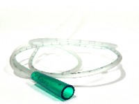
 So, in conclusion, we feel that it is both helpful to the gastroenterologist, and more importantly, beneficial for the patient to place a NG tube and perform a lavage. The following should be reported to the gastroenterologist when you call them:
- Was bloody material spontaneously returned upon placement of the tube
- What was the color of the material that was lavaged, bright blood red, maroon, clear with coffee-ground specks, etc. Please be cognizant of recent ingestion of blood-appearing food/drink by the patient
- If you find evidence of bleeding, please lavage at least 1-2 liters and tell us if the gastric contents clear of the bloody contents.
- If there is no evidence of blood in the gastric contents, please continue to lavage until you see bilious material returned, so the gastroenterologist can be sure that you are sampling contents beyond the pylorus, a common site of peptic ulcers.
8 comments on “To Lavage or Not to Lavage?”
In my opinion this patient does not need an NGT. He needs to be endoscoped regardless of the results of the NG aspirate. The timing of the endoscopy should be determined by an assessment as to the likelihood that his bleeding is life threatening. Clinically since he is hemodynamically stable he can be scoped within the next 24 hrs. The insertion of an NGT is quite an uncomfortable procedure and not nearly as well tolerated as an EGD.
I think you will invariably miss some patients that have frank blood sitting in the stomach or duodenum, who may have bad outcomes if not scoped sooner rather than later.
Neither patients nor doc like doing NG tubes, but isn’t getting a clear or bloody lavage pretty helpful in making the decision to scope now or later?
I think not. There are criteria which do not involve passing an NGT which can give an idea as to the rapidity with which the EGD should be done.The Rockall score, as well as others, look at the presence of comorbidity, hypotension that does not respone to a bolus of fluid, etc. If I came to the ER with melena I would refuse an NGT and simply ask for appropriate reususcitation and an EGD. Incidentally, it is usually the least experienced member of the team (medical student) who is given the job of passing an NGT without adequate supervision.
For more on the Rockall score- see pg. 3 of these British endoscopy guidelines:
http://www.bsg.org.uk/pdf_word_docs/nonvar3.pdf
There are certainly some instances where passing an NGT would not be of diagnostic value. Some examples would be in a patient admitted with hematemesis who has a known history of esophageal varices or a hemodynamically stable patient who presents with hematochezia. However, many gastroenterologists still utilize the NG lavage as a diagnostic and traige tool because it has been shown in several (though not all) studies to be a prognostic factor for clinical outcomes.
Of note, the complete Rockall score requires endoscopy as part of its scoring system and therefore cannot be used as an initial diagnostic tool. It has, post-endoscopy, been shown to be an effective predictor of rebleeding and all-cause mortality. There is an abbreviated Rockall score that does not require endoscopic findings but that scoring system has not been as extensively evaluated, as far as I am aware.
I’m an ED doc and had a terrible experience passing an NG tube down a potential upper GI bleed. So much so that it has me hesitant to perform it again. We passed the tube on a patient with melena and lavaged. Fluid came out clear so we promptly removed the tube. It is my belief that while we were extracting the tube we clipped a varice. Long story short the patient hemorrhaged and had a poor outcome. I have since read several studies that state to insert an NG tube even in the presence of varices, but I must say I’m very hesitant.
I’m also an ED doc and believe that the “triage” as to whether or not a patient should be scoped immediately or within 24 hours is purely a clinical decision based on observation of hemodynamic stability. A negative NG lavage in an unstable patient with suspected Upper GI bleed does not rule out a bleed passed a closed pyloric sphincter, and a positive NG lavage (that clears) in a stable patient does not warrant more urgent EGD than an unstable patient. It is common in my experience for GI docs to use lack of NGT, or negative or equivocal lavages as a way of delaying consultation till the morning.
Another ED doc weighs in: it seems to me that the issue is not what to do with a patient with a positive lavage. A positive lavage–ie either frank blood or significant coffee grounds in the lavage fluid–is no mystery. Get GI and hope they don’t request a bleeding scan (which takes forever at my institution and is not a good study for an remotely unstable patient). The issue is what to do with a negative lavage. Does anybody know the LR+ and LR- of lavage? If the pretest probability is high enough–which it usually is if you are even bothering to pass an NGT, shouldn’t egd be the next step anyway? and quickly? i recently had an UGIB go bad when GI refused to scope even though the pt came in unstable. he got appropriate resuscitation in the ED, stabilized, was almost rejected from the ICU because he initially refused NGT (he had a history of UGIB), eventually caved and had a negative lavage. GI decided to put off the scope till the morning–the patient continued to bleed overnight in the ICU and ended up with an antrectomy. I felt vindicated but it was very upsetting. thank goodness, the patient ultimately did well. but it was a huge mess.
also, i’d argue that distinguishing an upper from a lower GI bleed using NGT can be difficult. i’ve seen plenty of “negative” lavages turn out to be just a stopped or slowed UGIB rather than a LGIB.
Comments are closed.