
 By Karen McCloskey, MD
Peer ReviewedÂ
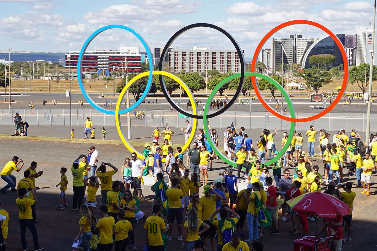
The world’s eyes were on Rio as the 2016 Summer Olympics wrapped up its first full week of competition. In the lap pool (fortunately not green, like the diving pool), United States swimmers had a fantastic games; Michael Phelps continued to demonstrate why he is the most decorated Olympian of all time, closing out his Olympic career with 28 medals, with 5 of his 23 gold medals earned in Rio (along with one silver).1 Katie Ledecky blew away the competition in women’s freestyle (finishing the 800m race nearly 12 seconds before the second-place winner), and Simone Manuel became the first African-American female swimmer to win an individual gold medal.
On land, Simone Biles stunned with her moves on the gymnastics floor, the uniforms of Egypt’s women’s beach volleyball team prompted discussions on culture, and Fiji’s rugby sevens side won the country’s first Olympic medal, and made it a gold.2
From awe-inspiring athletic achievement, we turn our attention to medicine, looking at some of this week’s most interesting literature:Â
Randomized Trial of Thymectomy in Myasthenia Gravis
About 10% of myasthenia gravis patients have thymomas that must be resected to prevent further spread of the disease3. Of the remaining patients, about 70% of patients have hyperplastic changes of the thymus that are not seen in healthy individuals4,5. Even in non-thymomatous myasthenia gravis patients, thymectomies have been performed for several decades, though rates of have declined over the last 15 years.6 Several retrospective studies and observational studies have been performed to examine the benefit of thymectomy in non-thymomatous myasthenia gravis, with promising, but inconsistent results. However, a systematic review was undertaken looking at 21 such studies, and it was determined that the amount of methodologic flaws in these studies precluded making a definitive recommendation based on those results.7
The Thymectomy Trial in Non-Thymomatous Myasthenia Gravis Patients Receiving Prednisone Therapy (MGTX) was published in the New England Journal of Medicine this week by Wolfe, et al. – it was an international randomized, single-blinded trial conducted by the National Institute of Neurological Disorders and Stroke (NINDS) to evaluate whether thymectomy combined with a standardized prednisone protocol was superior to prednisone therapy alone.8 The end points were lessening of myasthenic weakness, lower total doses of prednisone (given the high potential for side effects with glucocorticoid therapy), and improved quality of life. Participants were recruited from 67 centers in 18 countries (about half in the United States). 6958 patients were screened, with 2126 ultimately randomized. Inclusion criteria required a duration of myasthenia gravis less than 5 years; age of 18 to 65 years; certain antibody levels, as well as Myasthenia Gravis Foundation of America clinical classification of II-IV (representing mild, moderate, and severe generalized disease without crisis). Participants were allowed to be taking anticholinesterase therapy with or without oral steroids. Relevant exclusion criteria were thymoma on imaging or immunotherapy (besides prednisone). Participants were randomized to prednisone-only versus thymectomy groups. Those undergoing thymectomy had median sternotomy resection of all mediastinal tissue that could contain gross or microscopic thymus.
The results showed that patients in the thymectomy did better on all measured outcomes: a lower Quantitative Myasthenia Gravis score (representing improvement in overall disease symptoms) 6.15 vs 8.99 for the prednisone-only group (p<0.001, 95% CI 0.47 to 5.22) and lower average required prednisone dose (44mg vs 60mg based on standardized, alternatve-day dosing, p<0.001,95% CI, 7 to 25). In terms of quality of life, participants in the thymectomy group had fewer adverse reactions due to immunosuppressive medications, and 9% vs 37% (p<0.001) rate of hospitalizations for exacerbations.
This study showed a clear benefit to thymectomy in non-thymomatous myasthenia gravis, in terms of clinical status, prednisone dose (as well as adverse effects of immunosuppressive medications) and rate of hospitalizations for myasthenia exacerbations. Though the procedure itself is substantial, requiring median sternotomy, it appears the long-term affects of thymectomy are very positive for patients with myasthenia gravis.
Risk of lymphoma in patients exposed to antitumour necrosis factor therapy: results from the British Society for Rheumatology Biologics Register for Rheumatoid Arthritis
Biological agents like the Tumor Necrosis Factor inhibitors revolutionized the treatment of autoimmune diseases such as RA when they were introduced in the 1990s9–11However, there have been some concerns that these medications, due to their suppression of a key portion of the inflammatory pathway, might increase the risk of lymphoma development in patients with rheumatoid arthritis (RA).12 RA patients already have increased lymphoma risk13,14 thought to be due to the inflammatory nature of their disease.15 Conversely, it is possible that TNF inhibitors may, by way of inflammatory suppression, reduce the lymphoma risk in RA patients.
In the current issue of Annals of Rheumatic Diseases, the British Society for Rheumatology (BSR) led by Mercer et al., conducted a prospective cohort study using the data from the British Society for Rheumatology Rheumatoid Arthritis Register (BSRBR-RA).16 This register was established in 2001 in order to track, principally, the association between TNF inhibitors and development of lymphoma. Patients were separated into two cohorts, one biologic naïve group and the other biologic non-naïve. In the UK, TNF-I use is restricted to patients with highly active disease (based on a standardized Disease Activity Score) despite treatment with at least two non-biological disease-modifying drugs (csDMARDs). Once patients were enrolled with the BSRBR-RA, based on data collected from rheumatologists, they were also flagged in the national cancer registries (National Health Service Information Centre or Northern Ireland Cancer Registry). These cancer registries notified the BSRBR-RA if enrolled patients had a history of cancer or developed cancer after being registered. Patients were then followed until death or self-withdrawal from the study. Patients were excluded if they had prior diagnosis of lymphoproliferative or myeloproliferative malignancy, or if they had exposure to biological agents prior to enrollment. Available biologic agents at the time of the study were etanercept (ETA), infliximab (INF), and adalimumab (ADA).
The results showed that the 3,367 biological-naïve patients (treated with csDMARDs only) and the 11,931 showed no significant difference in the rate of lymphoma over the median 6.5 years of follow-up time. The results were calculated in patient-years with 154 cases per 100,000 (95% CI=104 to 220) in the 19,473 patient-years of the csDMARD group versus 88 cases per 100,000 (95% CI=70 to 109) in the biological group. When adjusted for confounders, the hazard ratio between these groups was 1.00 (95% CI 0.56 to 1.80). Each individual TNF-I was also evaluated and none demonstrated either increased or decreased risk of lymphoma.
This study provides a large-scale demonstration that there appears to be no increased risk of lymphoma when using TNF-I, helping rheumatologists feel safe in prescribing these very effective medications. Previous studies have shown mixed results in regard to this effect.17,18The large enrollment numbers and the advantage of the high percentage of disease capture due to the nature of the national registries give credence to the robustness of this study. The two main drawbacks of this study were insufficient power to detect the risk of individual subtypes of lymphoma, and the relatively short monitoring time, with the study tracking only up to 5 years after treatment initiation. This may not be a long enough period of treatment to detect medication-associated cancer risk. Further research studying larger populations, and with longer follow-up time, is needed to determine long-term lymphoma risk using these biological agents.
Association Between Occupational Exposures and Sarcoidosis: An Analysis From Death Certificates in the United States, 1988-1999
Mortality related to sarcoidosis has been increasing over the past several decades.19,20While the etiology of sarcoidosis has not been fully elucidated yet, the prevailing theory is that some environmental or occupational exposure triggers the disease in genetically susceptible individuals.21,22 Prior studies have examined the relationship between sarcoidosis and certain, select occupations (eg, firefighters23, navy recruits24, etc.), however no study has looked at the association of sarcoidosis using lifetime occupational data.
A study published in Chest this week by Liu et al, aimed to determine the association between sarcoidosis mortality and occupational exposures.25 Some of the basis of this study was to determine if previously studied gender differences in sarcoidosis rates (women are affected more frequently than men26) could be attributable to occupational disparities, or if occupational exposures imposed greater risk on women than men. These same questions were addressed taking into account racial differences as well.
The study used death certificate data from 1988 through 1999 as compiled by the National Center for Health Statistics from the 25 states that included Occupational Classification Codes during the studied time period. The researchers analyzed this data for exposures and job titles that had been found in prior research to have an association to sarcoidosis.
A total of 7,118,535 patients over age 14 found 3,393 sarcoidosis-related deaths, with an even ratio of men to women; however, sarcoidosis-related mortality was higher for woman than men with an adjusted mortality odds ratio (MOR) of 2.25 (95% CI, 2.10-2.42).  The study also found a sarcoidosis-related MOR of 1.52 of any occupational exposure versus no occupational exposure (95% CI, 1.35-1.71), with an increased risk for women with exposure vs women without (MOR 1.65, 95% CI 1.45-1.89). Implicated occupations were metal working, health care, teaching, sales, banking and administration. For certain occupations, the sarcoidosis-related mortality risks were high for blacks vs whites as well (ie, sarcoidosis-related deaths in healthcare fields had an MOR of 10.73 in black versus white subjects, 95% CI, 7.03-12.46). The study postulates that occupations related to “possible inhalational exposures†were associated with greater sarcoidosis-related mortality in men, while occupations related to “person-to-person contact†were associated with sarcoidosis-related mortality in women, and that this may correlate to previous findings that women are more prone to skin and eye involvement (contact-susceptible organs), while men are more prone to cardiac and pulmonary involvement (inhalation-suspectible organs)27–29Race differences are potentially related to genetic susceptibility.
While this study helps advance the research pointing to occupational-related sarcoidosis by its scale and breadth, it does leave some questions unanswered. Presumably, this study uses the term “no exposure†to mean occupations not previously shown to have sarcoidosis-related exposure risk; however, it could also mean subjects for whom no occupational data was available on death certificates – the manuscript is not clear on this point. In the former scenario, the presumption that all previously unstudied occupations pose no increased risk of sarcoidosis leaves the data open to confounding by occupations that have increased risk, but have not yet been identified in the literature. In the unlikely latter scenario, comparisons will have been made to a completely undefined control group. However, the study does advance the sarcoidosis-related research with regards to occupational exposure, which helps direct future research attempting to elucidate mechanisms of disease. It may also allow for better occupational safety standards in affected occupations.
Effect of a Cerebral Protection Device on Brain Lesions Following Transcatheter Aortic Valve Implantation in Patients With Severe Aortic Stenosis: The CLEAN-TAVI Randomized Clinical Trial
Transcatheter aortic valve implantation (TAVI) has allowed many high-risk patients the opportunity for a less-invasive procedure than an open surgical approach, with recent data suggesting a mortality benefit in TAVI patients.30 Unfortunately, stroke risk in aortic valve replacement patients, whether using surgical or transcatheter approaches, remains high – as many as 80% of TAVI patients are found to have new ischemic lesions on diffusion-weighted MRI31,32Several devices designed to prevent embolic debris from reaching the brain have been developed, with trends showing decreased number of ischemic lesions when these devices are used, however none have shown statistical significance.33
JAMA this week published results from the Claret Embolic Protection and TAVI (CLEAN-TAVI) from the University of Leipzig, a single-center, blinded, randomized control trial of 100 patients, to determine the efficacy of the Claret Montage Dual Filter System for cerebral protection.34 The Leipzig Heart Center, where this study was conducted, received a grants from Claret Medical as well as Medtronic (replacement valves were Medtronic CoreValves). The study included patients with severe, symptomatic aortic stenosis who were considered high-risk for surgical valve replacement. Exclusion criteria were pre-existing pacemaker, stroke within the previous 12 months, significant carotid, right subclavian or braciocephalic trunk stenosis, or anatomy unsuitable for TAVI.
Baseline MRI was compared to MRI at days 2 and 7 post-procedure. The primary end point was reduction in the number of new post-procedure on day 2 diffusion-weighted MRI (DWMRI). The group hypothesized that the cerebral protection device would lead to 50% reduction in new lesions on day 2 DWMRI in the regions protected by the cerebral protection filter. Results showed that TAVI was associated with DWMRI-positive brain lesions in 98% of patients in both the filter and the control groups, however the number of lesions was lower in the filter group (4.00 versus 10.00, difference 5.00, interquartile range 2.00-8.00, P<0.001). Secondary analysis showed that new lesion volume in the filter group was 242 mm3 (95% CI 159-353mm3, significantly lower than the 527mm3 (95% CI, 364-830) in the control group (difference of 234mm3, 95% CI, 91-406, P=0.001).  In both groups, 5 patients demonstrated neurologic symptoms of stroke, though all were minor and non-disabling.
While this study demonstrates a lower radiologic stroke burden in patients using this cerebral protection filter, it does not clearly show a clinical benefit to the use of this device. An equal number of patients in both groups showed neurologic manifestations of stroke, and all were minor. This study shows that there is some promise in the use of cerebral protection devices to reduce the stroke rate in TAVI, but a great deal more research is needed to demonstrate any clinical benefit before these devices become part of standard practice.
Mini-Cuts:
For the first time, a joint statement was published for the treatment of Type 2 Diabetes that included indications for when to recommend or consider bariatric surgery for patients as part of treatment for the disease.35
In a post-hoc analysis of the RE-LY trial (which looked at dabigtran versus warfarin in non-valvular atrial fibrillation) found that for patients with valvular heart disease (excluded by the original study) dabigitran carried a lower stroke risk and lower risk of significant bleeding as compared to warfarin.36
Finally, the US Preventive Services Task Force released guidelines on screening for dyslipidemia in young adults, aged 21-39, which showed that no study has examined the effects of lipid screening versus no screening in this age group, and therefore the Task Force is unable to recommend for or against screening at this time.37
Dr. Karen McCloskey is a 1st year resident at NYU Langone Medical Center
Peer reviewed by Kevin Hauck, MD, associate Editor, Clinical Correlations
Image courtesy of Wikimedia Commons
ReferencesÂ
- Michael Phelps: How swimming legend regained his “immortality.†http://edition.cnn.com/2016/08/13/sport/michael-phelps-legacy-rio-2016-olympics/.
- No Title. https://www.theguardian.com/artanddesign/2016/aug/12/twenty-olympic-moments-from-the-first-week-of-the-rio-games.
- Drachman DB. Myasthenia Gravis. N Engl J Med. 1994;330(25):1797-1810. doi:10.1056/NEJM199406233302507. http://www.ncbi.nlm.nih.gov/pubmed/8190158
- Conti-Fine B, Diethelm-Okita B, Ostlie N, Wang W, Milani M. Immunopathogenesis of myasthenia gravis. In: Kaminski H, ed. Myasthenia Gravis and Related Disorders. New York: Humana Press; 2009:43-70.
- Berrih-Aknin S, Le Panse R. Myasthenia gravis: A comprehensive review of immune dysregulation and etiological mechanisms. J Autoimmun. 2014;52:90-100. doi:10.1016/j.jaut.2013.12.011.
- Alshekhlee A, Miles JD, Katirji B, Preston DC, Kaminski HJ. Incidence and mortality rates of myasthenia gravis and myasthenic crisis in US hospitals. Neurology. 2009;72(18):1548-1554. doi:10.1212/WNL.0b013e3181a41211.
- Gronseth GS, Barohn RJ. Practice parameter: thymectomy for autoimmune myasthenia gravis (an evidence-based review): report of the Quality Standards Subcommittee of the American Academy of Neurology. Neurology. 2000;55(1):7-15. http://www.ncbi.nlm.nih.gov/pubmed/10891896
- Wolfe GI, Kaminski HJ, Aban IB, et al. Randomized Trial of Thymectomy in Myasthenia Gravis. N Engl J Med. 2016;375(6):511-522. doi:10.1056/NEJMoa1602489.
- Maini R, St Clair EW, Breedveld F, et al. Infliximab (chimeric anti-tumour necrosis factor alpha monoclonal antibody) versus placebo in rheumatoid arthritis patients receiving concomitant methotrexate: a randomised phase III trial. ATTRACT Study Group. Lancet. 1999;354(9194):1932-1939. doi:http://dx.doi.org/10.1016/S0140-6736(99)05246-0.
- Weinblatt ME, Keystone EC, Furst DE, et al. Adalimumab, a fully human anti-tumor necrosis factor alpha monoclonal antibody, for the treatment of rheumatoid arthritis in patients taking concomitant methotrexate: the ARMADA trial. Arthritis Rheum. 2003;48(1):35-45. doi:10.1002/art.10697.
- Klareskog L, Van Der Heijde D, De Jager JP, et al. Therapeutic effect of the combination of etanercept and methotrexate compared with each treatment alone in patients with rheumatoid arthritis: Double-blind randomised controlled trial. Lancet. 2004;363(9410):675-681. doi:10.1016/S0140-6736(04)15640-7.
- Brown SL, Greene MH, Gershon SK, Edwards ET, Braun MM. Tumor necrosis factor antagonist therapy and lymphoma development: Twenty-six cases reported to the Food and Drug Administration. Arthritis Rheum. 2002;46(12):3151-3158. doi:10.1002/art.10679.
- Isomäki HA, Hakulinen T, Joutsenlahti U. Excess risk of lymphomas, leukemia and myeloma in patients with rheumatoid arthritis. J Chronic Dis. 1978;31(11):691-696. doi:10.1016/0021-9681(78)90071-1. http://www.ncbi.nlm.nih.gov/pubmed/730824
- Smitten AL, Simon T a, Hochberg MC, Suissa S. A meta-analysis of the incidence of malignancy in adult patients with rheumatoid arthritis. Arthritis Res Ther. 2008;10(2):R45. doi:10.1186/ar2404.
- Baecklund E, Iliadou A, Askling J, et al. Association of chronic inflammation, not its treatment, with increased lymphoma risk in rheumatoid arthritis. Arthritis Rheum. 2006;54(3):692-701. doi:10.1002/art.21675.
- Mercer LK, Galloway JB, Lunt M, et al. Risk of lymphoma in patients exposed to antitumour necrosis factor therapy: results from the British Society for Rheumatology Biologics Register for Rheumatoid Arthritis. Ann Rheum Dis . 2016. doi:10.1136/annrheumdis-2016-209389 .
- Asten P, Barrett J, Symmons D. Risk of developing certain malignancies is related to duration of immunosuppressive drug exposure in patients with rheumatic diseases. J Rheumatol. 1999;26(8):1705-1714. http://www.ncbi.nlm.nih.gov/pubmed/10451066
- Askling J, Baecklund E, Granath F, et al. Anti-tumour necrosis factor therapy in rheumatoid arthritis and risk of malignant lymphomas: relative risks and time trends in the Swedish Biologics Register. Ann Rheum Dis. 2009;68(April):648-653. doi:10.1136/ard.2007.085852.
- Swigris JJ, Olson AL, Huie TJ, et al. Sarcoidosis-related mortality in the United States from 1988 to 2007. Am J Respir Crit Care Med. 2011;183(11):1524-1530. doi:10.1164/rccm.201010-1679OC.
- Hanley A, Hubbard RB, Navaratnam V. Mortality trends in asbestosis, extrinsic allergic alveolitis and sarcoidosis in England and Wales. Respir Med. 2011;105(9):1373-1379. doi:10.1016/j.rmed.2011.05.008.
- Valeyre D, Prasse A, Nunes H, Uzunhan Y, Brillet PY, M??ller-Quernheim J. Sarcoidosis. In: The Lancet.Vol 383.; 2014:1155-1167. doi:10.1016/S0140-6736(13)60680-7.
- Smith A, Brownell I, Sanchez M, Prystowsky S. Advances in the genetics of sarcoidosis. Clin Genet. 2008;73(5):401-412. doi:10.1111/j.1399-0004.2008.00970.x.
- Prezant DJ, Dhala A, Goldstein A, et al. The incidence, prevalence, and severity of sarcoidosis in New York City firefighters. Chest. 1999;116(5):1183-1193. doi:10.1378/chest.116.5.1183.
- Gorham ED, Garland CF, Garland FC, Kaiser K, Travis WD, Centeno JA. Trends and occupational associations in incidence of hospitalized pulmonary sarcoidosis and other lung diseases in Navy personnel: A 27-year historical prospective study, 1975-2001. Chest. 2004;126(5):1431-1438. doi:10.1378/chest.126.5.1431.
- Liu H, Patel D, Welch AM, et al. Association Between Occupational Exposures and Sarcoidosis. Chest. 2016;150(2):289-298. doi:http://dx.doi.org/10.1016/j.chest.2016.01.020.
- Birnbaum AD, Rifkin LM. Sarcoidosis: sex-dependent variations in presentation and management. J Ophthalmol. 2014;2014:236905. doi:10.1155/2014/236905.
- Judson M a, Boan a D, Lackland DT. The clinical course of sarcoidosis: presentation, diagnosis, and treatment in a large white and black cohort in the United States. Sarcoidosis Vasc Diffuse Lung Dis. 2012;29(2):119-127. http://www.ncbi.nlm.nih.gov/pubmed/23461074.
- Yanardaǧ H, Nuri Pamuk Ö, Karayel T. Cutaneous involvement in sarcoidosis: Analysis of the features in 170 patients. Respir Med. 2003;97(8):978-982. doi:10.1016/S0954-6111(03)00127-6.
- Grunewald J, Eklund A. Sex-specific manifestations of L??fgren’s syndrome. Am J Respir Crit Care Med. 2007;175(1):40-44. doi:10.1164/rccm.200608-1197OC.
- Adams DH, Popma JJ, Reardon MJ, et al. Transcatheter aortic-valve replacement with a self-expanding prosthesis. N Engl J Med. 2014;370(19):1790-1798. doi:10.1056/NEJMoa1400590.
- Fairbairn T a., Mather a. N, Bijsterveld P, et al. Diffusion-weighted MRI determined cerebral embolic infarction following transcatheter aortic valve implantation: assessment of predictive risk factors and the relationship to subsequent health status. Heart. 2012;98(1):18-23. doi:10.1136/heartjnl-2011-300065.
- Kahlert P, Knipp SC, Schlamann M, et al. Silent and apparent cerebral ischemia after percutaneous transfemoral aortic valve implantation: A diffusion-weighted magnetic resonance imaging study. Circulation. 2010;121(7):870-878. doi:10.1161/CIRCULATIONAHA.109.855866.
- Lansky AJ, Schofer J, Tchetche D, et al. A prospective randomized evaluation of the TriGuardTM HDH embolic DEFLECTion device during transcatheter aortic valve implantation: Results from the DEFLECT III trial. Eur Heart J. 2015;36(31):2070-2078. doi:10.1093/eurheartj/ehv191.
- Haussig S, Mangner N, MG D, al et. Effect of a cerebral protection device on brain lesions following transcatheter aortic valve implantation in patients with severe aortic stenosis: The clean-tavi randomized clinical trial. JAMA. 2016;316(6):592-601. http://dx.doi.org/10.1001/jama.2016.10302.
- Moncada R, Landecho MF, Fruhbeck G. Metabolic Surgery Enters the T2DM Treatment Algorithm. Trends Endocrinol Metab. 2016. doi:10.1016/j.tem.2016.07.006.
- Ezekowitz MD, Nagarakanti R, Noack H, et al. Comparison of Dabigatran versus Warfarin in Patients with Atrial Fibrillation and Valvular Heart Disease: The RE-LY Trial. Circulation. 2016. http://circ.ahajournals.org/content/early/2016/08/05/CIRCULATIONAHA.115.020950.abstract.
- Chou R, Dana T, Blazina I, Daeges M, Bougatsos C, Jeanne TL. Screening for Dyslipidemia in Younger Adults: A Systematic Review for the U.S. Preventive Services Task ForceScreening for Dyslipidemia in Younger Adults. Ann Intern Med. 2016;N/A(N/A):N/A – N/A. http://dx.doi.org/10.7326/M16-0946.