
Posted by Vivian Hayashi MD and Robert Smith MD, Mystery Quiz Section Editors
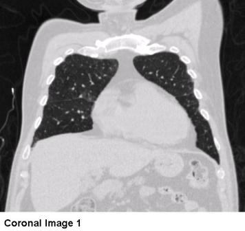
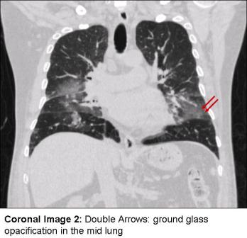
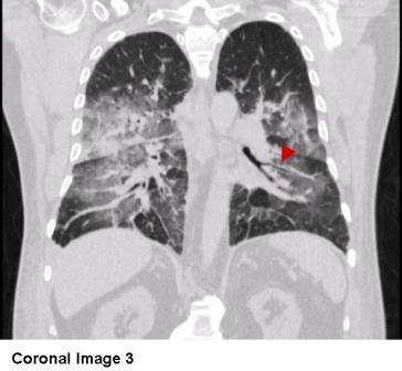
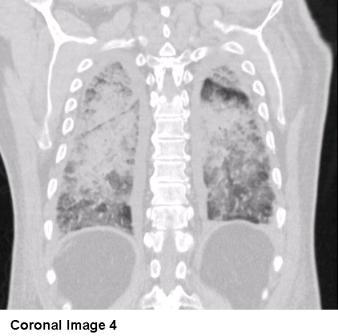
The answer to the mystery quiz is heart failure. The CXR shows bibasilar opacities with hilar fullness on the right. The CT images are remarkable for bilateral effusions, with dependent opacities that increase in density along the anterior-posterior axis. The lung appears clear in the anterior zone (Image 5, arrow; Coronal Image 1); ground glass opacification, characterized by parenchymal haziness which does not obscure the underlying pulmonary vessels, is evident in the mid lung (Image 5, double arrows; Coronal Image 2); and consolidation with air bronchograms is evident in the posterior areas (Image 5, arrowhead; Coronal Image 3). The coronal images show the same increase in density along the anterior-posterior axis (Coronal Images 1-4). Although the same density gradient of parenchymal fluid may be seen in non-cardiogenic pulmonary edema, the presence of bilateral effusions makes cardiogenic edema much more likely.
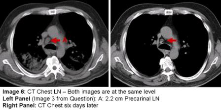
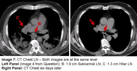
The mediastinal windows demonstrate adenopathy in precarinal, subcarinal and hilar areas (Image 6A, Image 7B and 7C, respectively). The fact that enlarged mediastinal and hilar lymph nodes may accompany congestive heart failure was not appreciated until relatively recently (Slanetz PJ et al. Mediastinal lymphadenopathy and hazy mediastinal fat: new CT findings of congestive heart failure. Am J Roentgenol 1998; 171: 1307-09).  Animal models indicate that enlarged nodes do not result simply from increased flow through interstitial lymphatics. An additional requirement is that the efferent draining lymphatic vessels empty into a vein that has an elevated pressure (>15cm H2O), as may occur in heart failure, but less likely to occur in cases of non-cardiogenic pulmonary edema. High venous pressures slow lymphatic outflow and contribute to proximal lymph node enlargement. The few reported biopsies of such nodes show an absence of inflammation, benign sinus histiocytosis, and slight follicular hyperplasia.
The CT images six days later show remarkable resolution of the airspace opacification after diuresis, along with a significant decrease in the lymphadenopathy (Images 6 and 7). We present this case to highlight an underappreciated cause of reversible lymphadenopathy and review the image findings of pulmonary edema, herein due to hypertensive heart disease.

One comment on “Mystery Quiz- The Answer”
That’s a great clinical pearl and something that can definitely be applied to my practice. Thanks for a new very interesting little piece of information.
Comments are closed.