
Posted By Robert Smith, MD Associate Professor of Medicine, Division Pulmonary and Critical Care Medicine
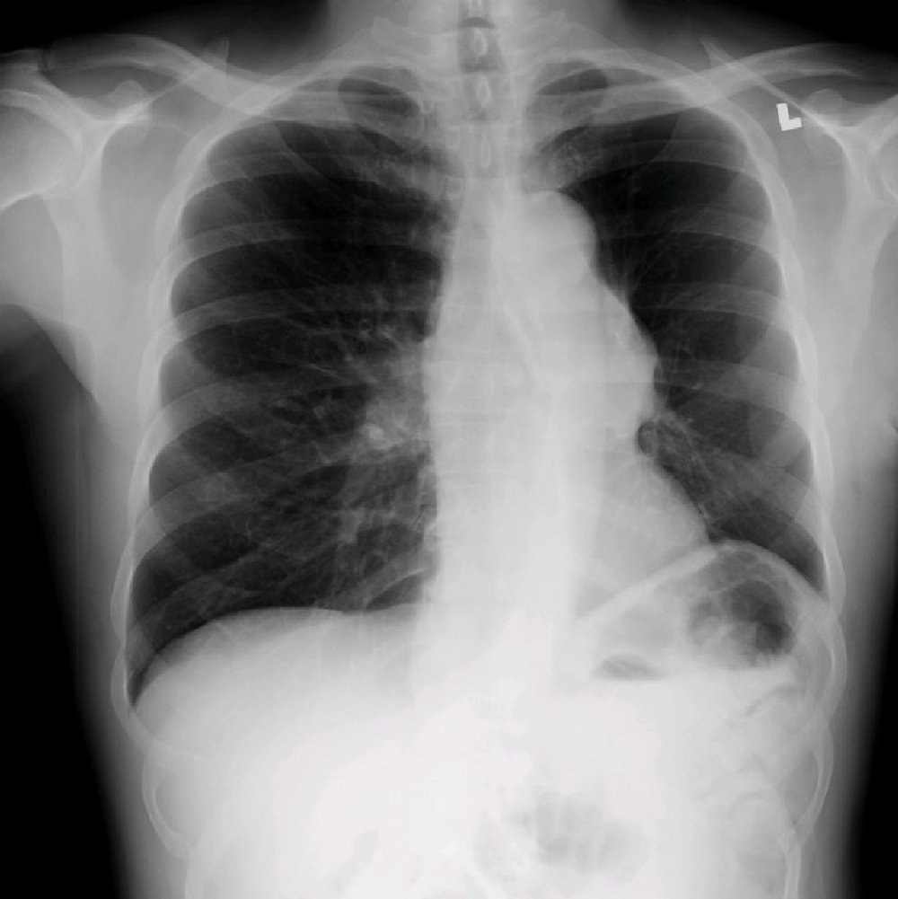
62 year old male smoker with past medical history notable for hypertension, copd, comes to clinic complaining of chronic cough x 6 months with minimal sputum, no wheezing. Lung exam shows decreased breath sounds throughout. Pt had the following CXR performed:
Click on Thumbnail to Enlarge
What's your diagnosis and the next step in his evaluation? Please submit your answers by clicking on the "comments" link below this post. For those of you who are unwilling to attach your name, you can post your comments anonymously. Responses will be posted for 1 week when the answer will be revealed…
25 comments on “Mystery Quiz-Radiology”
I would perform a CT Chest, wwo contrast.
My presumptive diagnosis is lung cancer, but I’m not intelligent enough to specify further.
It looks to me like an aspirated foreign body obstructing the left mainstem bronchus, with resulting volume loss on left–a peanut, based on its shape? I have new respect for radiologists!!!
Looks like a Type A (DeBakey Type II) dissection to me. Appropriate next step would be a spiral CT of the chest with contrast, or, if stable, an MRI or a TEE.
Agree with a foreign body aspiration…a tooth! I would perform Bronchoscopy and add to my collection!
I’m with Brabeck… enlarged aortic knob, trachea displaced to the right, chronic cough in a smoker with hypertension. CT with con or MRA.
I think there is an aortic aneurysm, but it may not yet have dissected given the lack of acute symptoms.
There seem to be multiple abnormalities on this chest XR:
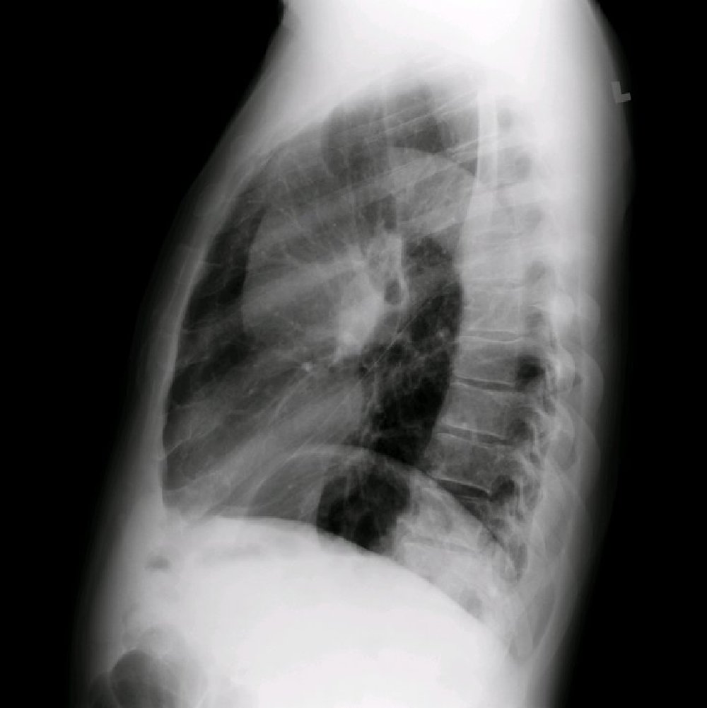
There is a collapse of part of the left lung. This is best seen on the lateral view. There are no obvious air-bronchograms in the density, c/w resorption (post-obstructive) atelectasis. This part of the lung seems to be the Left lower lobe. There are several reasons I think this finding is on the left. First, on the lateral view the minor fissure is going through the lesion. Second, part of the lesion obliterates the border of the L hemi-diaphragm on the lateral view (silhouette sign). Third, there is a relative volume loss on the left, with left hemidiaphragm elevation and mediastinal shift to the left.
In general, whenever a mediastinal shift is present, there is either something pulling it to the side of the shift (minus volume) or pushing on it from the other side (plus volume). In this case I believe that there is a minus volume on the left.
More importantly, there is a large left hilar mass. The left main bronchus and the carina are displaced to the right. Left main bronchus lumen is narrowed and seems somewhat irregular. Left upper pulmonary artery may be compromised with “amputation†at the hilum and relative oligaemia in the left upper lung field.
Aortic knob is normal, but ascending aorta is enlarged (as seen on the lateral). The heat shadow is displaced to the left, but otherwise seems normal.
As mentioned, there is an elevation of the left hemi-diaphragm. This is seen in the context of h/o COPD and a clear flattening of the right hemidiaphragm. When seen in association with LLL collapse, two possibilities are possible: impaired mobility of the hemidiaphragm (phrenic nerve paralysis) may result in decrease clearance of secretions in the LLL and plugging. Alternatively, the collapse could be a result of bronchial obstruction (intrinsic or extrinsic). In such a case hemi-diaphragm will get elevated simply because of decreased intrathoracic pressure. Importantly the first scenario does not cause mediastinal shifts, as seen here.
In summary, my DDx is that of a hilar mass. Theoretically, aortic root aneurysm can result in all these compressions. However, the abnormalities seem too extensive. I favor a diagnosis of left lung CA with hilar lymphadenopathy.
Finally, chronic cough may be caused by multiple mechanisms in this case, including irritation of the recurrent laryngeal nerve on the left (under the aortic arch) by the mass.
The next test should be CT of the chest with iv contrast.
Let’s see if I have made a complete ass of myself.
Clearly there is a mass in the left hilum, specifically there is a lesion in the middle/posterior mediastinum. As Greg Mints points out, the left hemidiaphragm is elevated. With the history of cough, I think this combination is suggestive of phrenic nerve involvement. A common diagnosis would be a neurogenic tumor, either arising from the phrenic or impinging the phrenic nerve.
If I am right, I thought of this by myself.
If I am wrong, Josh Olstein told me to write this.
emphysema
widened aorta, also with left lower lobe atelectasis
would perform ct chest w iv contrast
I am neither a Dr or pulmonologist but do have several lung conditions which I have done extensive research on. My guess would be COPD and/or lung cancer of sorts. Note the extensive mass on the left lung at the bottom and the small node on the right lung, suggestive of having been there a while or spreading rapidly. Further tests with contrast and possibly a biopsy. What is the correct diagnosis??? Eager to learn more.
I think this patient developed pulmonary embolism (Thrombo embolic diseases) lead to volume loss on left side and widen pulmonary artery (westermark sign)
VOLUME LOSS IN LEFT LUNG FIELD WITH HYPERINFLATION IN RT LUNG FIELD WITH MILD DEVIATION OF MEDIASTINUM TOWORDS LT SIDE
HILD DENSITY NOTED ON LT SIDE AND ALSO RT PARATRACHEAL REGION
POSSIBLY A LT HILAR MASS(?NODE) CAUSING OBSTUCTION TO LT MAJOR BRONCHUS WITH COMPENSATORY CHANGES IN RT LUNG
RML collapse with Left middle mediastinal, paratracheal mass probably lymph node or parenchymal mass.
CT followed by brochoscopy with biopsy and sputum analysis.
Smoking history + signs of hyperinflation = Emphysema
Smoking is a strong risk factor for central lung cancer (Squamous and Small cell cancers)
A Lt Hilar mass is noted pushing the lower trachea to the right as well as forming a silhouette with the adjacent Cardiac shadow denoting a contigous location (Mediastinal or lower lobe which would be supported by its location above the oblique fissure in lateral view)
The left hemidiaphragm is elevated than the right (opposite is normal) pointing to a Lt phrenic nerve involvement by the hilar pathology
I would recommend a Chest CT scan with IV contrast
Agree with Dr. Brabeck and others… there is an enlarged Aortic Root, volume loss on left in the setting of extensive COPD–without back or substernal pain it is less likely to be an acute dissection- but may be growing and compressing left main steam bronchus.. and causing cough..CT with contrast… tread carefully with CV surg…
there is widening of superior mediastinum , in lateral view widening is cintinuous with aorta. DD – strong posibility of aortic aneurysm, with AR which lead to LV failure. aortic dissection and AR may be.
Volume loss on the left. Compression of left main bronchus. Abnormal mediastinum on the left. Small left pulmonary artery. Vascular abnormaluity involving pulmonary conus likely. CT or pulmonary angio
PA-hyperinflation from COPD, mediastinal shift to L , raised L hemidiaphragm
but clues for collapse not obvious? enlarge aortic knob without calcification & cardiomegaly, reason for shift unlikely
Lat- enlarged hilum, blunted retrocardial border, incidental osteoporosis, no additional clue of any lytic bone lesion.
? CA lung
next- CT/bronchoscopy
Bronchogenic carcinoma of the mainstem left bronchus and bilateral hilar congestion
The pa and lateral cxr shows features of a chronic obstructive airway disease as evidenced by compensatory emphysema of rt lung causing mediastinal shift to lt side,the flattening of rt diaphragm is part of the compensatory mechanism. the left hilar shadow with convex lateral margin is the prominent main pulmonary artery, there is generalised pulmonary oligaemia with poor broncohvascular markings..probable of pulmonary hyper tension.In the rt hilum there is a wedge opacity -westmarc-sign probably due to a pulmonary embolism. The probable diagnosis is pulmonary embolism in a pre existing chronic pulmonary disease back ground with features of pulmonary hypertension. further evaluation with CT CHEST CONRAST STUDY AND CT PULMONARY ANGIOGRAM AND VENTILATION /PERFUSION SCAN FOR LUNG.
I’m still learning. Likely left hilar mass causing collapse of left lower lobe with compensatory hyperinflation of right lung.
PROBABLY COPD with severe pulmonary hypertension with ortner”s sign
chronic obstructive air way disease with unfolding of arch of aorta due to hypertention.
i would order a ct for mediastinal evaluation
this patient has prominent right hillum and traction of diaphraghm,that due to smoking ,malignancy is the best diagnosis that must be rule out. I recommend broncoscopy to this patient.
Comments are closed.