Peer Reviewed
Mr. Q is a 55-year-old male smoker who presents with recurrent chest pain in the mornings over the past several months. The patient reports being awakened from sleep at approximately 5:00 a.m. each morning with the same diffuse chest “pressure.” The pain typically lasts on the order of minutes, resolves, and then recurs at five-minute intervals in the same fashion for a total duration of two hours. The pain always occurs at rest and is never precipitated by exertion or emotional stress. The chest pain is generally associated with a sense of palpitations and occasional dizziness and light-headedness. An exercise stress test showed good exercise capacity without ST segment changes, even at target heart rate. Given the history, a diagnosis of coronary artery spasm was suggested. The patient was given a trial of diltiazem therapy, with marked improvement in his chest pain episodes thereafter.
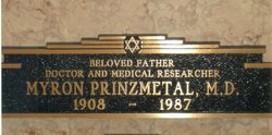
In his landmark article in 1959, Dr. Myron Prinzmetal described a distinct type of “variant angina,” termed Prinzmetal’s angina. This chest pain tended to occur at rest (i.e. was not associated with increased cardiac work), waxed and waned cyclically, occurred at the same time each day, and could be accompanied by arrhythmias including ventricular ectopy, ventricular tachycardia, ventricular fibrillation, and various forms of AV block [1]. The patient’s EKG during painful episodes typically showed ST segment elevations (occasionally accompanied by reciprocal ST depressions), whereas the EKG obtained after the pain had resolved showed resolution of these ST segment changes [1]. Prinzmetal postulated that this separate clinical entity was due to transient spasm (“increased tonus”) of a large arteriosclerotic artery, causing temporary transmural ischemia in the distribution supplied by that artery.
It is important to note that, although ST elevation would be diagnostic, it is frequently not observed in cases of coronary artery spasm. Rather, the diagnosis of coronary artery spasm should be suspected based on the timing of chest pain and the presence of syncope, arrhythmia or cardiac arrest.
It was subsequently demonstrated that such episodes of coronary artery spasm can occur not only in patients with underlying fixed coronary artery obstruction but also in patients whose coronary arteries are anatomically normal [2-7]. Selzer et al. actually compared the syndromes of coronary artery spasm between nine patients with anatomically normal coronary arteries and 20 patients with obstructive coronary lesions [8]. Selzer et al. found that the non-coronary artery disease (CAD) group of patients was more likely to have a long history of nonexertional angina without prior infarction, normal EKG at rest with ST elevations in the inferior leads, conduction disease, and bradyarrhythmias during episodes of arterial spasm. Conversely, the CAD group of patients was more likely to have prior “effort angina” and prior infarction, as well as ST elevation in the anterolateral leads, ventricular ectopy and ventricular tachyarrhythmias.
Castello et al. also compared the syndromes of coronary artery spasm in 77 patients with underlying CAD (fixed coronary stenosis greater than or equal to 50%) and 35 patients with normal or minimally diseased coronary arteries [4]. These authors found, similarly, that angina exclusively at rest tends to occur in patients with structurally normal coronary arteries and that these patients tended to have more diffuse coronary artery spasms affecting more than one artery. In contrast, patients with underlying CAD usually had more focal coronary artery spasms superimposed on their fixed stenotic lesions.
The question arises as to what could be triggering coronary artery spasm in patients with structurally normal coronary arteries? As Prinzmetal suggested, “the distinctive dissimilarities [between typical angina and variant angina] are due to profound physiological and chemical rather than anatomical differences” [1]. These physiological and chemical differences are multi-factorial. Kugiyama et al. demonstrated that there is a deficiency in endothelial nitric oxide (NO) bioactivity in Prinzmetal’s angina-prone arteries; this defect makes those arteries especially sensitive to the vasodilator effect of nitroglycerin and the vasoconstrictor effect of acetylcholine [9]. Miyao et al. used intravascular ultrasound to show that Prinzmetal’s angina patients had diffuse intimal thickening of their coronary arteries, despite an angiographically normal appearance. This intimal hyperplasia was thought to be mediated by deficient NO activity [10]. NO is involved in the regulation of basal vascular tone and helps to mediate flow-dependent vasodilation, as well as suppressing the production of endothelin-1 and angiotensin-II, both of which are powerful vasoconstrictors [11]. As a result of all of these effects, deficient endothelial NO activity predisposes to coronary artery spasm. Endothelial NO is made by the endothelial NOS (e-NOS) gene, which has been found to have many genetic polymorphisms associated with coronary artery spasm [11]. It is important to note, however, that endothelial NO synthase polymorphisms are found in only one-third of patients with coronary spasm; accordingly, other genes or factors are most likely involved [11].
In a review article, Kusama et al. [12] highlighted several additional pathophysiologic contributors to Prinzmetal’s angina, including enhanced vascular smooth muscle contractility mediated by the Rho/Rho-kinase pathway [13-14], elevated markers of oxidative stress [11,15], low-grade chronic inflammation [11], and cigarette smoking [11,15] in addition to genetic polymorphisms of endothelial NO synthase (NOS) [11,15]. Polymorpisms of various genes may explain the higher incidence of Prinzmetal’s angina in the Japanese population as compared to the Caucasian population [12].
As our understanding of the pathophysiology behind Prinzmetal’s angina has evolved, new ways of diagnosing Prinzmetal’s angina have emerged. These diagnostic maneuvers typically involve provoking episodes of Prinzmetal’s angina under controlled settings (e.g. during coronary angiography) with acetylcholine, ergonovine, hyperventilation, and cold pressor stress testing. Okumura et al. showed that intracoronary injection of acetylcholine could be reliably used to induce coronary artery spasm with 99% specificity [16], a conclusion further supported by Miwa et al. [17]. Ergonovine, an ergot alkaloid and alpha-agonist that causes vasoconstriction, can similarly be used to induce episodes of coronary artery spasm accompanied by the characteristic chest pain and EKG changes that occur during spontaneous episodes of Prinzmetal’s angina [18-19]. Song et al. suggested ergonovine echocardiography as an effective screening test for coronary artery spasm, even before coronary angiography, with a sensitivity of 91% and a specificity of 88% [20]. Subsequent studies found that this was indeed an effective, safe, and well-tolerated screening test for coronary artery spasm [21-22].
It is important to note that provocation of arterial spasm with acetylcholine or ergonovine confers a multitude of risks including arrhythmias, hypertension, hypotension, abdominal cramps, nausea, and vomiting [11]. More serious complications include ventricular fibrillation, myocardial infarction, and death [23,24]. Quantitative estimates of the risks incurred by such invasive testing are on the order of 1% [25,26]. In one study, serious major complications, such as sustained ventricular tachycardia, shock, and cardiac tamponade occurred in four out of 715 patients (0.56%) receiving provocative acetylcholine testing [25]. In another study, nine patients out of 921 (1%) had more minor complications (nonsustained ventricular tachycardia [n=1], fast paroxysmal atrial fibrillation [n=1], symptomatic bradycardia [n=6], and catheter-induced spasm [n=1]) after undergoing acetylcholine provocation testing [26]. While such invasive testing is generally considered a safe technique to assess coronary vasomotor dynamics, these maneuvers should only be performed by qualified physicians in carefully controlled settings, where the patient may be properly and quickly resuscitated as needed [11].
Testing a different diagnostic strategy, Hirano et al. noted that a diagnostic algorithm of hyperventilation for six minutes, followed by cold water pressor for two minutes under continuous EKG and echocardiographic monitoring had a 90% Sensitivity, 90% specificity, 95% positive predictive value, and 82% negative predictive value for diagnosing vasospastic angina [27]. The combination of respiratory alkalosis from the hyperventilation as well as the reflex sympathetic coronary vasoconstriction in response to the cold pressor test [28], together, helped to induce coronary artery spasm and diagnose Prinzmetal’s angina. More recently, Hwang et al. suggested that measuring the change in coronary flow velocity of the distal left anterior descending artery (LAD) via transthoracic echo during the cold pressor test may provide additional diagnostic utility, with a sensitivity of 93.5% and a specificity of 82.4% for diagnosing coronary artery spasm [29].
In an article published in JACC in 2013, the Japanese Coronary Spasm Association (JCSA) discussed a comprehensive clinical risk score to aid in prognostic stratification of patients with coronary artery spasm [30]. A multicenter registry study of 1429 patients, median age 66 years, with a median follow-up period of 32 months, was performed. The primary endpoint was defined as major adverse cardiac events (MACE), including cardiac death, nonfatal myocardial infarction, hospitalization due to unstable angina pectoris, heart failure, and appropriate ICD shocks during the follow-up period that began at the date of the diagnosis of coronary artery spasm. In particular, cardiac death, nonfatal myocardial infarction and ICD shocks were categorized as hard MACE. The secondary endpoint was all-cause mortality. The study identified seven predictors of MACE: history of out-of-hospital cardiac arrest (4 points), smoking, angina at rest alone, organic coronary stenosis, multivessel spasm (2 points each), ST segment elevation during angina and beta-blocker use (1 point each). Based on total score, three risk categories were defined: low risk (score of 0 to 2, which included 598 patients), intermediate risk (score of 3 to 5, which included 639 patients) and high risk (score of 6 or more, which included 192 patients). The incidences of major adverse cardiac events in the low-, intermediate-, and high-risk patients were 2.5%, 7.0%, and 13.0%, respectively (p<0.001). This scoring system, known as the JCSA risk score, may help provide a comprehensive risk assessment and prognostic stratification scheme for patients with coronary artery spasm.
In terms of treatment, calcium channel blockers (e.g. nifedipine, diltiazem and verapamil) are the mainstay of therapy for coronary artery spasm. The goal of such therapy is to prevent vasoconstriction and promote coronary artery vasodilation. In one study of 245 patients with coronary artery spasm who were followed for an average of 80.5 months, the use of a calcium cannel blocker therapy was an independent predictor of myocardial-infarct-free survival in patients with coronary artery spasm [31]. In another observational study of 300 patients with coronary artery spasm, calcium channel blockers were effective in alleviating symptoms in over 90-percent of patients [32]. The drugs were evaluated and ranked as follows: markedly effective, leading to complete elimination of angina attacks within 2 days; effective, leading to complete elimination of attacks after 2 days or a reduction in the number of attacks to less than half during the periods of drug administration in the hospital; ineffective, leading to no reduction to less than half during the periods of drug administration. Efficacy rates (including markedly effective as well as effective categories) for nifedipine, diltiazem and verapamil were 94.0%, 90.8%, and 85.7%, respectively. Rarely, cases are refractory to medical therapy and literature exists to support the effectiveness of surgical revascularization in these circumstances [33].
It is clear that the phenomenon of “variant angina” is a complicated, multifaceted product of forces that are not only anatomical, but also genetic, chemical, physiological and behavioral in nature. While endothelial nitric oxide bioactivity appears to play a critical role in this process, there are undoubtedly several other factors involved. Over time, our knowledge of the pathophysiology driving Prinzemetal’s angina will continue to expand, as will our diagnostic and therapeutic repertoire for this fascinating clinical entity.
Dr. Anjali Varma Desai is a 3rd year resident at NYU Langone Medical Center
Peer Reviewed by Harmony R. Reynolds, MD, Medicine (Cardio Div), NYU Langone Medical Center
References:
1. Prinzmetal M, Kennamer R, Merliss R, Wada T, Bor N. Angina pectoris: I: a variant form of angina pectoris: preliminary report. Am J Med. 1959; 27: 375–388 http://www.ncbi.nlm.nih.gov/pubmed/14434946
2. Maseri A, Severi S, Nes MD, et al. “Variant” angina: one aspect of a continuous spectrum of vasospastic myocardial ischemia. Pathogenetic mechanisms, estimated incidence and clinical and coronary arteriographic findings in 138 patients. Am J Cardiol. Dec 1978;42(6):1019-35 http://www.ncbi.nlm.nih.gov/pubmed/727129
3. Cheng TO, Bashour R, Kelser GA Jr, et al: Variant angina of Prinzmetal with normal coronary arteriograms: a variant of the variant. Circulation 1973; 47: 476-485. http://circ.ahajournals.org/content/47/3/476.abstract
4. Castello R, Alegria E, Merino A, Soria F, Martinez-Caro D. Syndrome of coronary artery spasm of normal coronary arteries: Clinical and angiographic features. Angiology 1988; 39: 8-15. http://www.ncbi.nlm.nih.gov/pubmed/3341608
5. Oliva PB, Potts DE, Pluss RG. Coronary arterial spasm in Prinzmetal angina: documentation by coronary arteriography. N Engl J Med 1973; 288: 745-751. http://www.ncbi.nlm.nih.gov/pubmed/4688712
6. Endo M, Kanda I, Hosoda S, et al. Prinzmetal’s variant form of angina pectoris: Re-evaluation of mechanisms. Circulation 1975; 52: 33-37. http://circ.ahajournals.org/content/52/1/33.abstract?cited-by=yes&legid=circulationaha;52/1/33&related-urls=yes&legid=circulationaha;52/1/33
7. Huckell VF, McLaughlin PR, Morch JE, Wigle ED, Adelman AG: Prinzmetal’s angina with documented coronary artery spasm: Treatment and follow-up. Br Heart J 1981 June; 45(6): 649-655. http://www.ncbi.nlm.nih.gov/pmc/articles/PMC482578/
8. Selzer A, Langston M, Ruggeroli C, et al: Clinical syndrome of variant angina with normal coronary arteriogram. N Engl J Med 1976; 295: 1343-1347. http://www.ncbi.nlm.nih.gov/pubmed/980080
9. Kugiyama K, Yasue H, Okumura K, et al. Nitric oxide activity is deficient in spasm arteries of patients with coronary spastic angina. Circulation 1996 Aug 1; 94(3): 266-71. http://www.ncbi.nlm.nih.gov/pubmed/8759065
10. Miyao Y, Kugiyama K, Kawano H, et al. Diffuse intimal thickening of coronary arteries in patients with coronary spastic angina. J Am Coll Cardiol. 2000 Aug; 36(2): 432-7. http://www.ncbi.nlm.nih.gov/pubmed/10933354
11. Yasue H, Nakagawa H, Itoh T, Harada E, Mizuno Y. Coronary artery spasm – clinical features, diagnosis, pathogenesis and treatment. J Cardiol 2008; 51: 2-17. http://www.ncbi.nlm.nih.gov/pubmed/18522770
12. Kusama Y, Kodani E, Nakagomi A, et al. Variant angina and coronary artery spasm: the clinical spectrum, pathophysiology and management. J Nihon Med Sch. 2011;78(1):4-12. Review. http://www.researchgate.net/publication/50351691_Variant_angina_and_coronary_artery_spasm_the_clinical_spectrum_pathophysiology_and_management
13. Shimokawa H, Seto M, Katsumata N, et al. Rho-kinase mediated pathway induces enhanced myosin light chain phosphorylations in a swine model of coronary artery spasm. Cardiovasc Res 1999; 43: 1029-1039. http://cardiovascres.oxfordjournals.org/content/43/4/1029.full
14. Masumoto A, Mohri M, Shimokawa H, et al. Suppression of coronary artery spasm by a Rho-kinase inhibitor fasudil in patients with vasospastic angina. Circulation 2002; 105: 1545-1547. http://circ.ahajournals.org/content/105/13/1545.abstract
15. Miwa K, Fujita M, Sasayama S. Recent insights into the mechanisms, predisposing factors and racial differences of coronary vasospasm. Heart Vessels 2005; 20: 1-7. http://www.ncbi.nlm.nih.gov/pubmed/15700195
16. Okumura K, Yasue H, Matsuyama K, et al. Sensitivity and specificity of intracoronary injection of acetylcholine for the induction of coronary artery spasm. J Am Coll Cardiol. 1988 Oct;12(4):883-8. http://www.unboundmedicine.com/evidence/ub/citation/3047196/Sensitivity_and_specificity_of_intracoronary_injection_of_acetylcholine_for_the_induction_of_coronary_artery_spasm_
17. Miwa K, Fujita M, Ejiri M, Sasayama S. Usefulness of intracoronary injection of acetylcholine as a provocative test for coronary artery spasm in patients with vasospastic angina. Heart Vessels. 1991;6(2):96-101 http://www.ncbi.nlm.nih.gov/pubmed/1906457
18. Schroeder JS, Bolen JL, Quint RA, et al. Provocation of coronary spasm with ergonovine maleate: new test with results in 57 patients undergoing coronary arteriography. Am J Cardiol 1977; 40: 487-491. http://www.ncbi.nlm.nih.gov/pubmed/910712
19. Heupler FA, Proudfit WL, Razavi M, et al. Ergonovine maleate provocative test for coronary arterial spasm. Am J Cardiol 1978; 41: 631-640. http://www.ncbi.nlm.nih.gov/pubmed/645566
20. Song JK, Lee SJ, Kang DH, Cheong SS, Hong MK, Kim JJ, Park SW, Park SJ. Ergonovine echocardiography as a screening test for diagnosis of vasospastic angina before coronary angiography. J Am Coll Cardiol. 1996 Apr;27(5):1156-61. http://www.ncbi.nlm.nih.gov/pubmed/8609335
21. Palinkas A, Picano E, Rodriguez O, et al. Safety of ergot stress echocardiography for non-invasive detection of coronary vasospasm. Coron Artery Dis 2001 Dec; 12(8): 649-54. http://www.ncbi.nlm.nih.gov/pubmed/11811330
22. Djordjevic-Dikic A, Varga A, Rodriguez O, et al. Safety of ergotamine-ergic pharmacologic stress echocardiography for vasospasm testing in the echo lab: 14 year experience on 478 tests in 464 patients. Cardiologia 1999 Oct; 44(10): 901-6. http://www.ncbi.nlm.nih.gov/pubmed/10630049
23. M. Nakamura, A. Takeshita, Y. Nose. Clinical characteristics associated with myocardial infarction, arrhythmias and sudden death in patients with vasospastic angina. Circulation 1987; 75: 1110-1116.
24. R.J. Myerburg, K.M. Kessler, S.M. Mallon, et al. Life-threatening ventricular arrhythmias in patients with silent myocardial ischemia due to coronary artery spasm. New England Journal of Medicine 1992; 326: 1451-1455.
25. Sueda S, Saeki H, Otani T, et al. Major complications during spasm provocation tests with an intracoronary injection of acetylcholine. Am J. Cardiol. 2000; 85(3): 391.
26. Ong P, Athanasiadis A, Borgulya G, et al. Clinical usefulness, angiographic characteristics, and safety evaluation of intracoronary acetylcholine provocation testing among 921 consecutive white patients with unobstructed coronary arteries. Circulation 2014; 129(17): 1723.
27. Hirano Y, Ozasa Y, Yamamoto T, et al. Diagnosis of vasospastic angina by hyperventilation and cold-pressor stress echocardiography: comparison to I-MIBG myocardial scintigraphy. J Am Soc Echocardiogr. 2002 Jun;15(6):617-23. http://www.unboundmedicine.com/washingtonmanual/ub/citation/12050603/Diagnosis_of_vasospastic_angina_by_hyperventilation_and_cold_pressor_stress_echocardiography:_comparison_to_I_MIBG_myocardial_scintigraphy_
28. Raizner AE, Chahine RA, Ishimori T, et al. Provocation of coronary artery spasm by the cold pressor test. Hemodynamic, arteriographic and quantitative angiographic observations. Circulation. 1980; 62: 925-932. http://circ.ahajournals.org/content/62/5/925.citation
29. Hwang HJ, Chung WB, Park JH, et al. Estimation of coronary flow velocity reserve using transthoracic Doppler echocardiography and cold pressor test might be useful for detecting of patients with variant angina. Echocardiography. 2010 Apr;27(4):435-41. http://www.ncbi.nlm.nih.gov/pubmed/20113325
30. Takagi Y, Takahashi J, Yasuda S, et al. Prognostic Stratification of Patients with Vasospastic Angina: A Comprehensive Clinical Risk Score Developed by the Japanese Coronary Spasm Association. JACC 2013; 62(13): 1144-1153.
31. Yasue H, Takizawa A, Nagao M, et al. Long-term prognosis for patients with variant angina and influential factors. Circulation. 1988;78(1):1.
32. Kimura E, Kishida H. Treatment of variant angina with drugs: a survey of 11 cardiology institutes in Japan. Circulation 1981 April; 63(4): 844-8.
33. Ono T, Ohashi T, Asakura T, Shin T. Internal mammary revascularization in patients with variant angina and normal coronary arteries. Interact Cardiovasc Thorac Surg. 2005;4:426–428.


One comment on “Unraveling The Mysteries of Prinzmetal’s Angina: What Is It And How Do We Diagnose It?”
Comments are closed.