
 Class act is a feature of Clinical Correlations written by NYU 3rd and 4th year medical students. Prior to publication, each commentary is thoroughly reviewed for content by a faculty member.
Commentary by Daniel Green MSIV and Boris Kobrinsky MD, Assistant Professor, NYU Division of Oncology
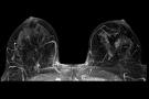
In 2008, an estimated 182,460 women in the United States will be diagnosed with invasive breast cancer, and 40,480 women will die of the disease as it remains the demographic’s second leading cause of cancer mortality.(1) Fortunately, breast cancer is one of the screenable cancers, and screening mammography has been shown to detect asymptomatic breast cancer at an early stage and reduce all-cause mortality when followed with appropriate treatment.(2,3) However, plain mammography has a sensitivity of about 85 percent, resulting in an estimated 21.8% of cases that are node positive at the time of diagnosis.(4)
The use of MRI in breast cancer screening is receiving increased attention, and the American Cancer Society has recently recommended it as an adjunct to plain mammography in the screening of high risk patients.(5) These patients include women with dense breast tissue, a personal history of breast cancer or lobular carcinoma in situ, prior mantle irradiation for Hodgkin’s lymphoma, and a strong family history of breast cancer.
Women with inherited BRCA1 and BRCA2 mutations have the greatest risk of breast cancer. Though only five to ten percent of women with breast cancer have one of the two mutations, those with a BRCA genotype have a lifetime risk of 65 to 80 percent of developing the disease.(5) This population tends to develop more aggressive breast cancers with significant risk of disease starting as early as age 30.
Several large, prospective, nonrandomized trials have been conducted to evaluate the use of MRI as an adjunct to plain mammography in screening high risk women for breast cancer. The largest of these studies, conducted in The Netherlands and published in 2004, evaluated both MRI and mammography in 1,909 high risk women.(6) The investigators found high sensitivity of MRI (80 percent) compared to that of mammography, whose sensitivity plummets to 33 percent in this high risk population. On the other hand, MRI had lower specificity than mammography (90 and 95 percent, respectively). Several other studies in North America and Europe have recapitulated these results.(7-11)
These studies were included in a recently published meta-analysis of 11 prospective studies on screening women at high risk of breast cancer with a combination of MRI and plain mammography. The investigators concluded that screening with mammography plus MRI may exclude breast cancer better then mammography alone in a population of women with a strong genetic predisposition to breast cancer.(12)
Enhancement of invasive breast carcinomas in contrast studies with gadolinium enables the increased sensitivity of MRI. However, many benign breast lesions enhance with gadolinium, resulting in a lower specificity. In women not characterized as high risk, the likelihood of false positives may lead to an unacceptable amount of recalls and biopsies. Because of the increased cancer rate in high risk women, the incidence of benign biopsy following MRI is similar to that of a population-based study using plain mammography.(13) In these patients, the benefit of high sensitivity MRI may outweigh the effects of lower specificity, though data on survival are not yet available.
A recent study from Stanford University evaluated the cost-effectiveness of supplementing screening mammography with MRI for carriers of BRCA mutations.(14) Health benefits were measured in terms of total health-related costs and quality-adjusted life years. The researchers found that screening MRI was more cost-effective in BRCA1-positive women compared to BRCA2-positive women because BRCA1 mutations confer a higher risk of breast cancer. However, they did not find that women with BRCA1 mutations aged 25-34 were at high enough risk to justify annual MRI screening. In addition, women with BRCA1 mutations over the age of 55 suffered from declining quality of life and competing risks for death, thus rejecting MRI as cost-effective for this age group. This leaves women with BRCA1 mutations aged 35-54 as the group most likely to benefit from MRI while taking cost into account.
MRI is also being utilized for screening of both the ipsilateral and contralateral breasts in women recently diagnosed with breast cancer. The prevalence of synchronous MRI detected breast cancer is considered to be between 1 and 9.5 percent, and these cancers are often both mammographically and clinically occult.(15,16) As is the case with standard breast cancer screening, the false positive rate is high due to limited specificity.
Current evidence suggests that MRI can benefit women at high risk, while there is much weaker evidence supporting its use in women of normal risk. Further research is necessary to develop the best method to improve screening in women at an intermediate level of risk, in whom the benefit of MRI remains unclear.
REFERENCES
1. Jemal A, Siegel R, Ward E, et al. Cancer statistics, 2008. CA Cancer J Clin 2008;58(2):71-96.
2. Nyström L, Andersson I, Bjurstam N, et al. Long-term effects of mammography screening: updated overview of the Swedish randomised trials. Lancet 2002;359(9310):909-19.
3. Glass AG, Lacey JV Jr, Carreon JD, Hoover RN. Breast cancer incidence, 1980-2006: combined roles of menopausal hormone therapy, screening mammography, and estrogen receptor status. J Natl Cancer Inst 2007;99(15):1152-61.
4. Weaver DL, Rosenberg RD, Barlow WE, et al. Pathologic findings from the Breast Cancer Surveillance Consortium: population-based outcomes in women undergoing biopsy after screening mammography. Cancer 2006;106(4):732-42.
5. Saslow D, Boetes C, Burke W, et al. American Cancer Society guidelines for breast screening with MRI as an adjunct to mammography. CA Cancer J Clin 2007;57:75-89.
6. Kriege M, Brekelmans CT, Boetes C, et al. Efficacy of MRI and mammography for breast cancer screening in women with a familial or genetic predisposition. N Engl J Med 2004;351:427-437.
7. Kuhl CK, Schrading S, Leutner CC, et al. Mammography, breast ultrasound, and magnetic resonance imaging for surveillance of women at high familial risk for breast cancer. J Clin Oncol 2005;23:8469-8476.
8. Leach MO, Boggis CR, Dixon AK, et al. Screening with magnetic resonance imaging and mammography of a UK population at high familial risk of breast cancer: a prospective multicentre cohort study (MARIBS). Lancet 2005;365:1769-1778.
9. Lehman CD, Blume JD, Weatherall P, et al. Screening women at high risk for breast cancer with mammography and magnetic resonance imaging. Cancer 2005;103:1898-1905.
10. Sardanelli F, Podo F. Breast MR imaging in women at high risk of breast cancer. Is something changing in early breast cancer detection? Eur Radiol 2007;17(4):873-87.
11. Warner E, Plewes DB, Hill KA, et al. Surveillance of BRCA1 and BRCA2 mutation carriers with magnetic resonance imaging, ultrasound, mammography, and clinical breast examination. JAMA 2004;292:1317-1325.
12. Warner E, Messersmith H, Causer P, et al. Systematic Review: Using magnetic resonance imaging to screen women at high risk for breast cancer. Ann Intern Med 2008;148:671-79.
13. Warren RM, Pointon L, Caines R, et al. What is the recall rate of breast MRI when used for screening asymptomatic women at high risk? Magn Reson Imaging 2002;20(7):557-65.
14. Plevritis SK, Kurian AW, Sigal BM, et al. Cost-effectiveness of screening BRCA1/2 mutation carriers with breast magnetic resonance imaging. JAMA 2006;295:2374-2384.
15. Lehman CD, Gatsonis C, Kuhl CK, et al. MRI evaluation of the contralateral breast in women with recently diagnosed breast cancer. N Engl J Med 2007;356(13):1295-303.
16. Lee SG, Orel SG, Woo IJ, et al. MR imaging screening of the contralateral breast in patients with newly diagnosed breast cancer: preliminary results. Radiology 2003;226(3):773-8.
3 comments on “Class Act: The Use of MRI in Breast Cancer Screening”
I have worked with medical imaging facilities for a number of years and have recently read several articles about the use of MRI for breast scans. This article is very educational; I did not realize that the breast MRI scan is best for high risk women. I have also read a number of articles about MRI being used for stress tests. I look forward to reading more about these advances.
Marge
http://www.remakehealth.com
I am a bit perplexed regarding some of the assertions made in this article. Specifically, it is suggested that the article by Saslow, et al. stated that women with a prior history of breast CA, dense breast tissue on mammography, and women found to have LCIS or ADH, constituted groups for which screening breast MRI was determined to be indicated, according to evidence-based analysis.
In their article which presented the current ACS guidelines, Saslow, et al. stated that evidence was insufficient to recommend for or against adjunctive MRI screening for women with, “. . . heterogeneously or extremely dense breast on mammography”, or for, “. . . a personal history of breast cancer, including ductal carcinoma in situ (DCIS)”. Similarly, women with LCIS, ALH and ADH were also cited as groups for whom the evidence was insufficient to recommend for or against MRI screening ans an adjunct to routine mammographic screening.
Saslow, et al. go on in their article to cite what appears to be an evolving body of evidence that suggests that there may be a role either for screening MRI presently on a case-by-case basis for some of these risk groups, or in the future on a more generalized basis following the generation of more experimental data, but to suggest that the current ACS Guidelines recommend routine MRI screening for all of these groups seems to be an overstatement.
Bob Mooney
I’m confused. As a invasive lobular Type II breast CA survivor post right mastectomy-is a MRI of the left breast recomended?
Comments are closed.